35 blood flow through the heart diagram
Heart is a vital organ that you cannot live without. The function of heart is quite complex, but you can understand things better through the heart diagram labeled below. It provides information about different chambers of the heart and valves that help transfer blood from one part of your heart to another. Heart is responsible for the blood flow to every single part of the body through constant contraction and relaxation of cardiac muscles. This is the reason why we hear rhythmic beats all the time. Heart pumps pure blood to different parts of the body and then takes the deoxygenated blood from all the parts to the lungs for oxygenation. Normally in a minute the heart beats 72 times and pumps ...
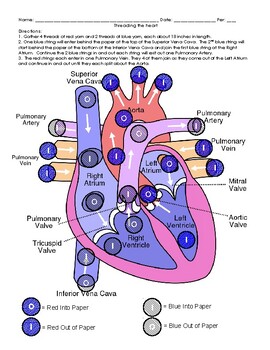
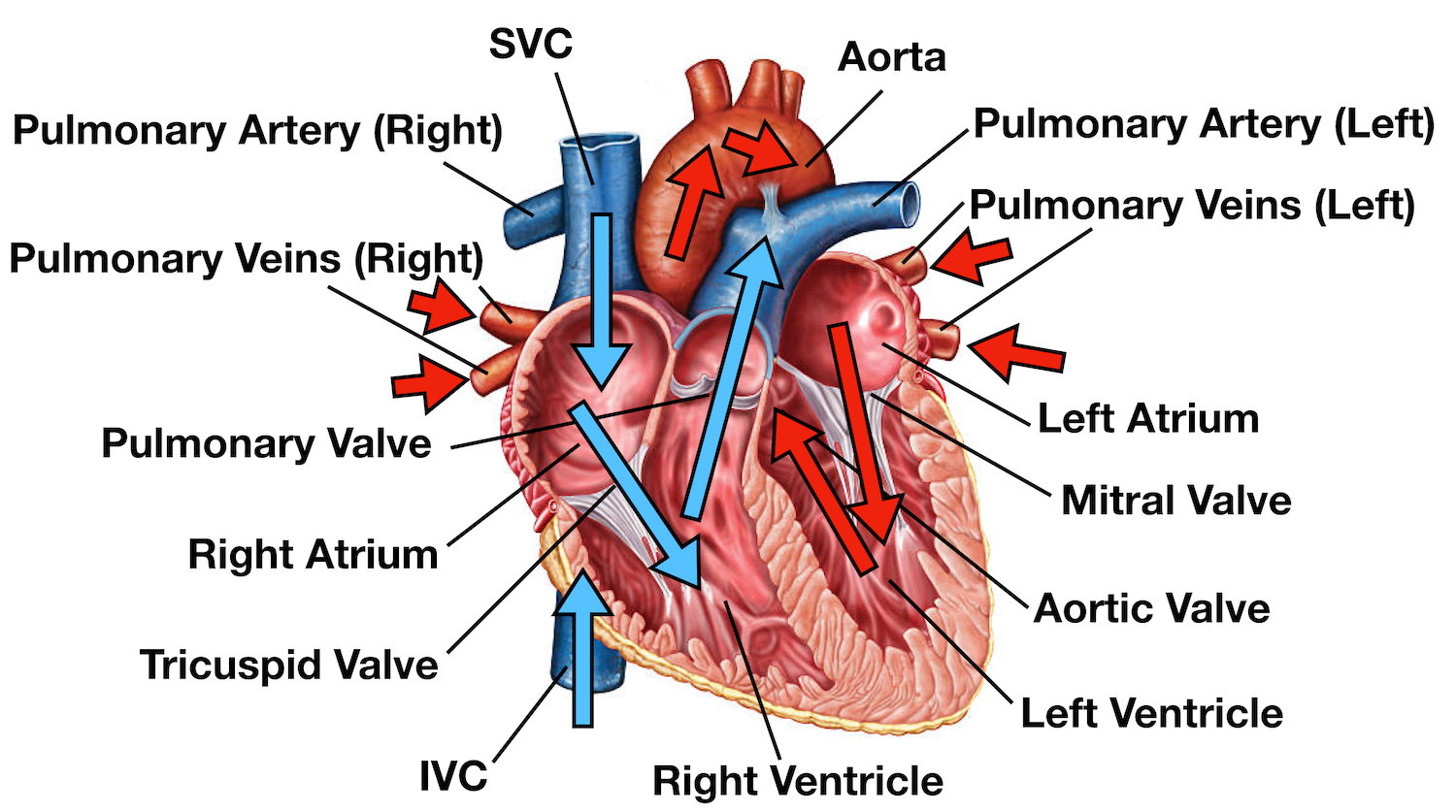
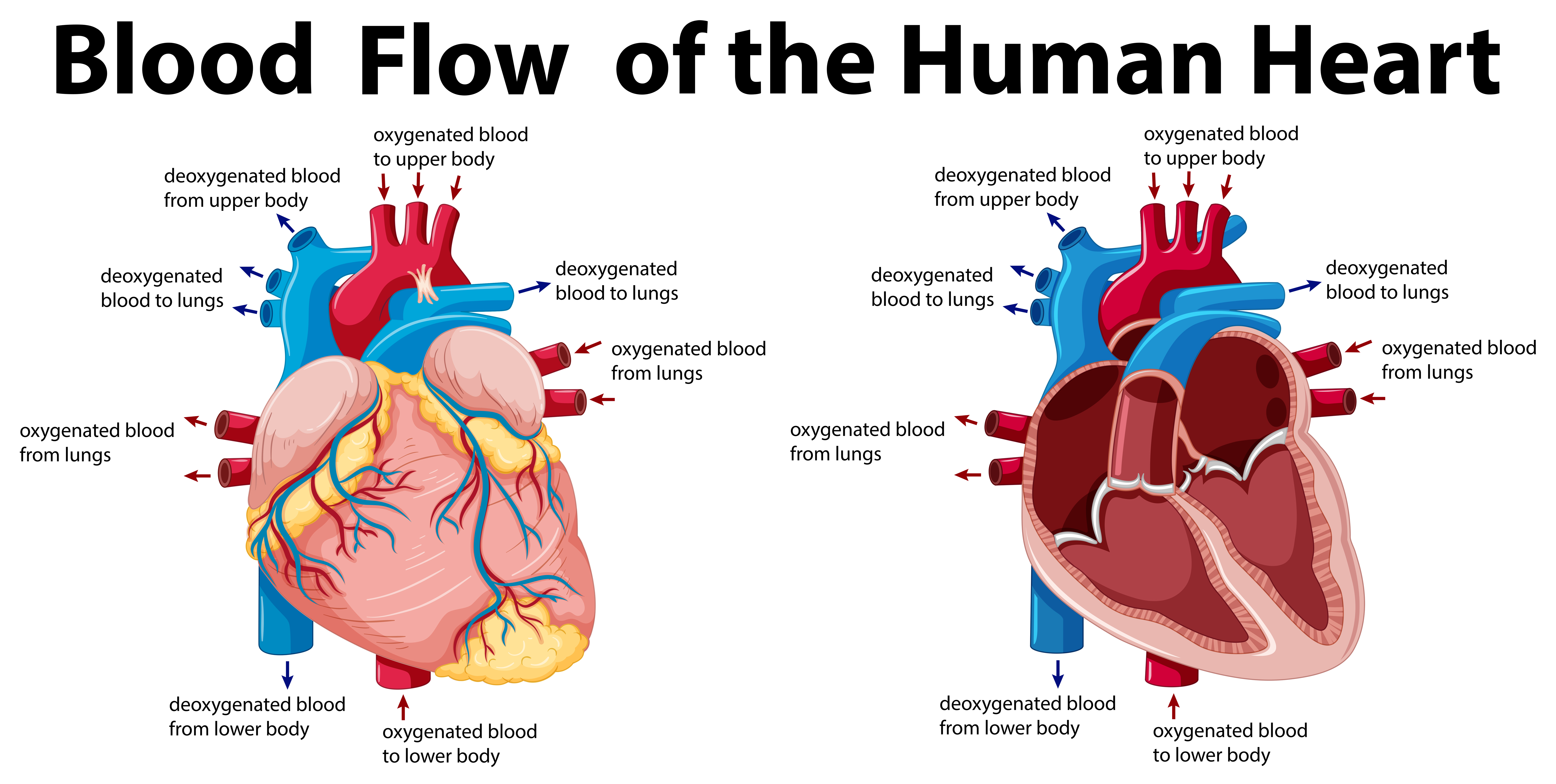
Blood enters the heart through two large veins, the inferior and superior vena cava, emptying oxygen-poor blood from the body into the right atrium of the heart ...What does the heart look like... · Heart diagram parts, location...

Blood flow through the heart diagram
Diagram of the human fetal circulatory system. The circulatory system, consisting of the heart and blood vessels, forms relatively early during embryonic development, and continues to develop in complexity within the growing fetus. A functional circulatory system is a biological necessity, since mammalian tissues can not grow more than a few cell layers thick without an active blood supply ... Congestive heart failure: The heart is either too weak or too stiff to effectively pump blood through the body. Shortness of breath and leg swelling are common symptoms. Cardiomyopathy: A disease ... Includes how blood flows through the heart and lungs, where the heart is located, what your heart and coronary arteries look like, and how the heart beats.
Blood flow through the heart diagram. 14+ Human Heart Blood Flow Diagram. This diagram shows how blood flows through a healthy heart. Heart blood flow heart flow learn biology heart anatomy cardiac nursing anatomy study circulatory system anatomy and physiology study human body: Circulatory - Biology 100 from austinlaulainen.weebly.com It is the only artery in the… A: om the body: t lung VEINS: trium VENTRICLE: o the lung TRIUM VE A: o the body: o the lungs APEX VENTRICLE: o the body all VE TRIUM VEINS: trium e e A: om the body 13 Aug 2020 — The heart contains four chambers, two atria and two ventricles. The blood that is returned to the right atrium is deoxygenated and s passed into ... Blood Flow Through the Heart. STUDY. Learn. Flashcards. Write. Spell. Test. PLAY. Match. Gravity. Created by. HPS3290 PLUS. Terms in this set (27) superior vena cava. receives blood from the head and arms and chest and empties into the right atrium of the heart. pulmonary artery.
Trace path of blood in diagram of fetal circulation (see diagram) Three shunts in the fetal circulation 1. Ductus arteriosus … protects lungs against circulatory overload … allows the right ventricle to strengthen … hi pulmonary vascular resistance, low pulmonary blood flow … carries mostly med oxygen saturated blood. 2. Ductus venosus … fetal blood vessel connecting the umbilical ... HCL Learning DigiSchool presents you animated study material on structure of the Human Heart. It describes the location, structure and function of the human ... Blood Flow Through the Heart. Beginning with the superior and inferior vena cavae and the coronary sinus, the flowchart below summarizes the flow of blood through the heart, including all arteries, veins, and valves that are passed along the way. 1. Superior and inferior vena cavae and the coronary sinus 2. Rt. atrium 3. Diagram of Blood Flow Through the Heart. You must have seen the human heart diagram. Thus, you will be aware of the anatomy of the human heart. If not, you can have a look at the labeled diagram of the human heart present in this article.
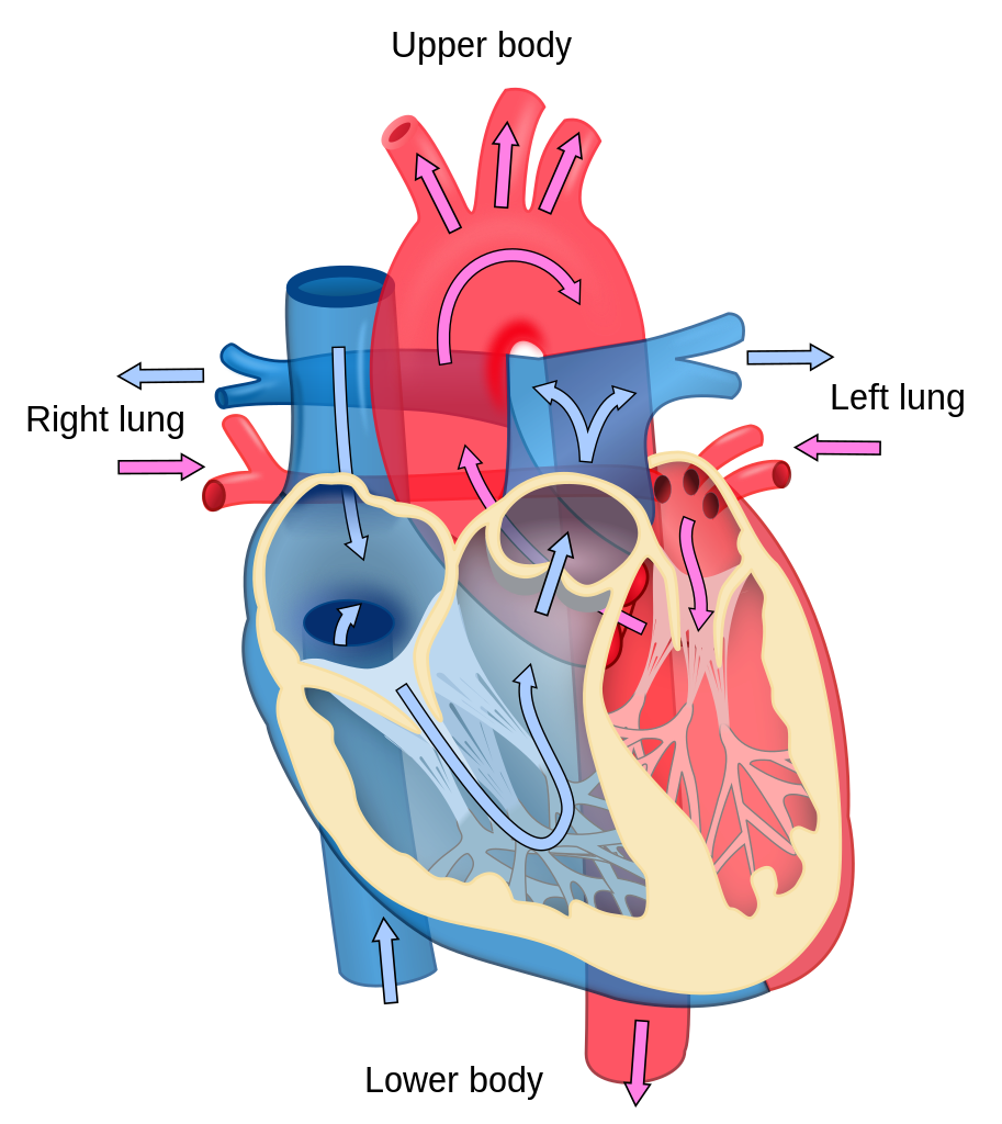
Blood flow through the heart made easy with a simple diagram of the cardiac circulation pathway and steps in order. Heart anatomy, video, quiz, and chart included! Great for USMLE, nursing, students, doctors, and medical learners. Trace the blood flow through the heart, and learn the names of important parts of the sequence. Try to follow the pathway of blood shown in Figure 1.4. The Pathway of Blood to and from the Heart 1. Blood that has circulated through the body, which has lost its oxygen and collected carbon dioxide, enters through the vena cava into the right atrium of the heart. 2. The right atrium contracts and ... allow blood to flow in only one direction through the heart chambers—from the atria through the ventricles and out the great arteries leaving the heart (see Figure 11.3b). The atrioventricular (AV) valves (a″tre-o-ven-trik′u-lar) are located between the atria and ventricles on each side. These valves prevent backflow into the atria when Blood leaves the heart through the aortic valve, into the aorta and to the body. This pattern is repeated, causing blood to flow continuously to the heart, lungs and body. How does blood flow through your lungs? Once blood travels through the pulmonic valve, it enters your lungs. This is called the pulmonary circulation.
5 A An Iconic Diagram For Blood Circulation In The Heart This Download Scientific Diagram
4 Feb 2021 — As the heart beats, it pumps blood through a system of blood vessels, called the circulatory system. The vessels are elastic tubes that carry ...
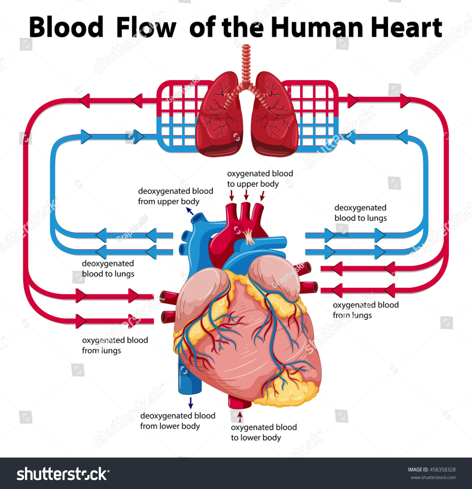
Diagram Showing Blood Flow Human Heart Stock Vector Royalty Free 458358328
Blood Flow Step by Step · The blood first enters the right atrium. · The blood then flows through the tricuspid valve into the right ventricle. · When the heart ...

Blood Flow Through The Heart Diagram Quizlet
Blood Flow Through the Heart study guide by Alesha_Jeter includes 8 questions covering vocabulary, terms and more. Quizlet flashcards, activities and games help you improve your grades.

How The Heart Works Diagram Anatomy Blood Flow
Includes how blood flows through the heart and lungs, where the heart is located, what your heart and coronary arteries look like, and how the heart beats.
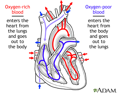
Circulation Of Blood Through The Heart Medlineplus Medical Encyclopedia Image
Congestive heart failure: The heart is either too weak or too stiff to effectively pump blood through the body. Shortness of breath and leg swelling are common symptoms. Cardiomyopathy: A disease ...

Review Labeled Heart Diagram Ppt Video Online Download
Diagram of the human fetal circulatory system. The circulatory system, consisting of the heart and blood vessels, forms relatively early during embryonic development, and continues to develop in complexity within the growing fetus. A functional circulatory system is a biological necessity, since mammalian tissues can not grow more than a few cell layers thick without an active blood supply ...
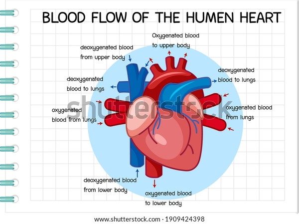
Vektor Stok Diagram Blood Flow Human Heart Illustration Tanpa Royalti 1909424398

Heart Blood Flow Video
14 Steps Of

The Heart And Circulation Of Blood

Pin On Blusas De Moda

Blood Flow Of Human Heart Diagram Stock Vector Illustration Of Health Girl 208659386

Coronary Circulation Anatomical Cross Section Diagram Labeled Vector Illustration Scheme Blood Flow Circuit From Body Through Heart And Lungs And Back To The Body Stock Illustration Download Image Now Istock
Blood Circulation In The Fetus And Newborn

Diagram Of Blood Flow Of The Human Heart Stock Vector Illustration Of Infographic Flow 208658709
Heart Diagram And Modeling Blood Flow Activity By Ruizscience Tpt

Diagram Showing Blood Flow Of The Human Heart Illustration Canstock

The Blood Flow Through The Heart Quiz
File Heart Diagram Blood Flow En Svg Wikipedia

Heart Ck 12 Foundation

3 Blood Flow Through The Heart 1 Download Scientific Diagram

Hand Drawn Illustration Of Human Heart Anatomy Educational Diagram Showing Blood Flow With Main Parts Labeled Vector Illustration Easy To Edit Royalty Free Cliparts Vectors And Stock Illustration Image 91670931

Blood Flow Human Heart Diagram Royalty Free Vector Image

Blood Circulation Through The Heart Download Scientific Diagram

Blood Flow Through The Heart Diagram Quizlet

File Heart Diagram En Svg Wikimedia Commons
Heart Blood Flow Simple Anatomy Diagram Cardiac Circulation Pathway Steps Ezmed
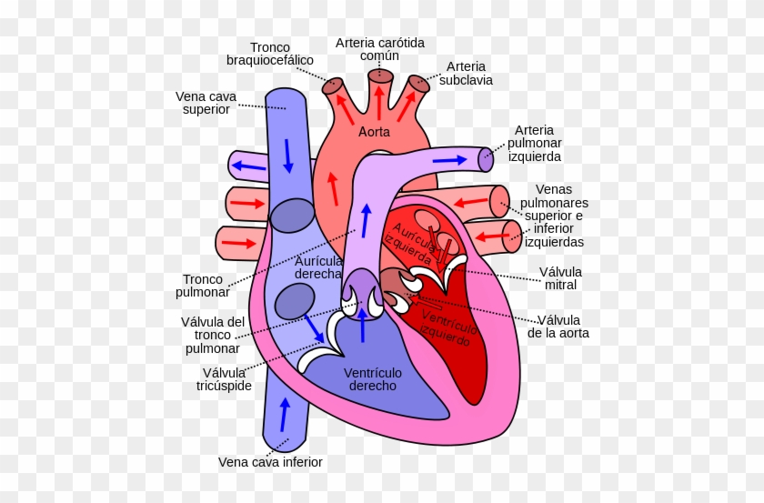
Diagram Of The Human Heart Flow Of Blood Through The Heart Free Transparent Png Clipart Images Download
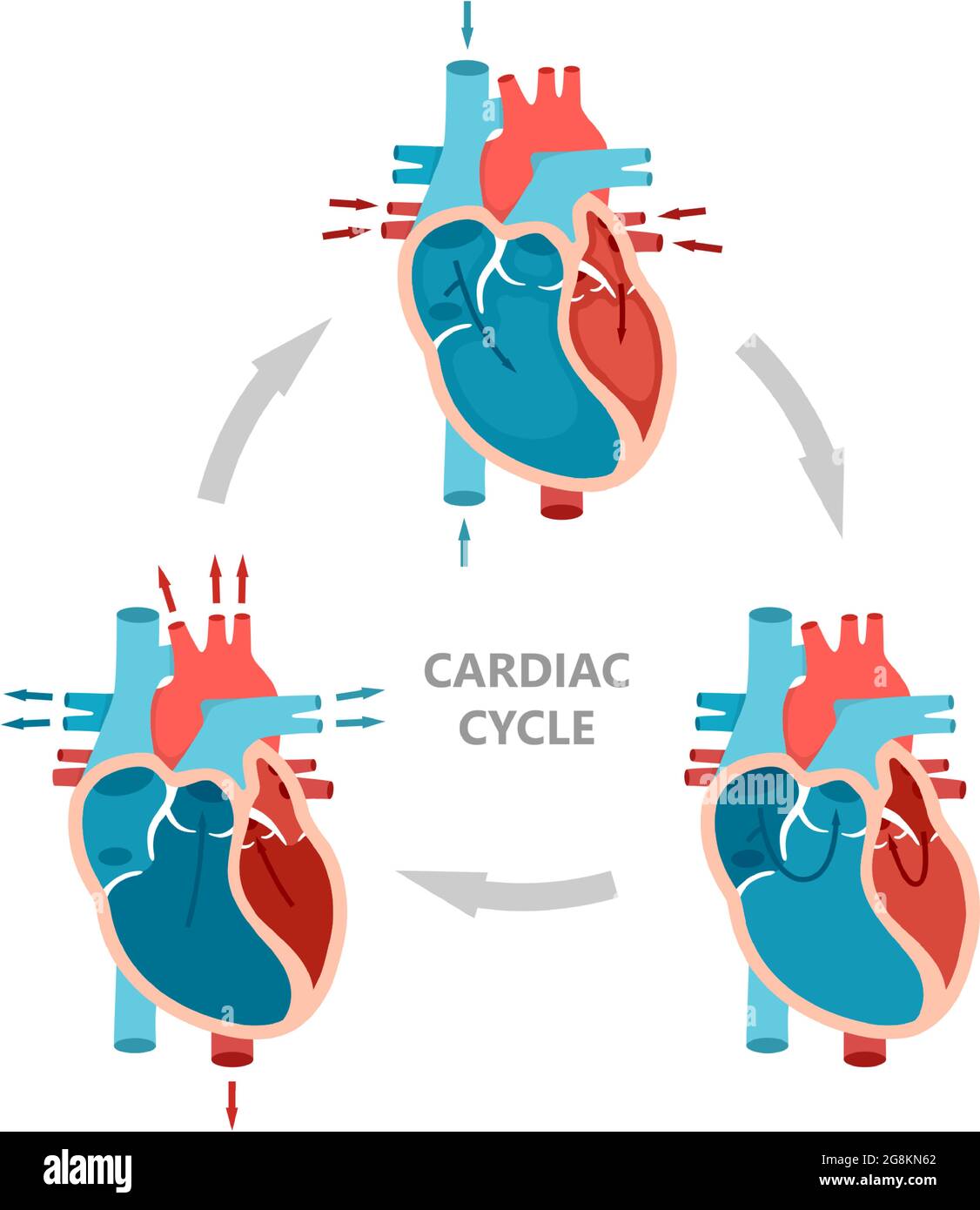
Phases Of The Cardiac Cycle Diastole Atrial Systole And Atrial Diastole Heart Anatomy Diagram With Blood Flow Stock Vector Image Art Alamy
Blood Flow Of The Human Heart 416577 Vector Art At Vecteezy
Anatomy And Physiology Of The Heart Normal Function Of The Heart Cardiology Teaching Package Practice Learning Division Of Nursing The University Of Nottingham

Diagram Showing Blood Flow In Human Heart Illustration Canstock

Cardio Respiratory System Heart Lungs Nurse Medical Knowledge Nursing Study
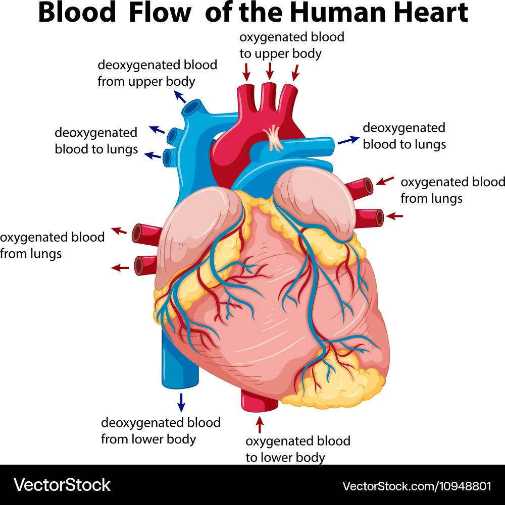
Diagram Showing Blood Flow In Human Heart Vector Image

Chart Showing Blood Flow Of Human Heart Royalty Free Vector
0 Response to "35 blood flow through the heart diagram"
Post a Comment