39 cow eye dissection diagram
Your eyes see, but how does vision happen? Find out how the eyes and brain work together in this eye video. Learn how to dissect a cow's eye in your classroom. This resource includes: a step-by-step, hints and tips, a cow eye primer, and a glossary of terms.
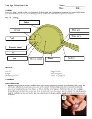
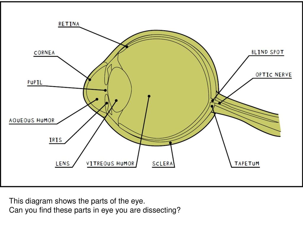
5. Use the diagram to identify the internal structures of the eye. 6. Remove the vitreous humor and lens from the front portion of the ...4 pages
Cow eye dissection diagram
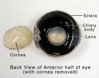
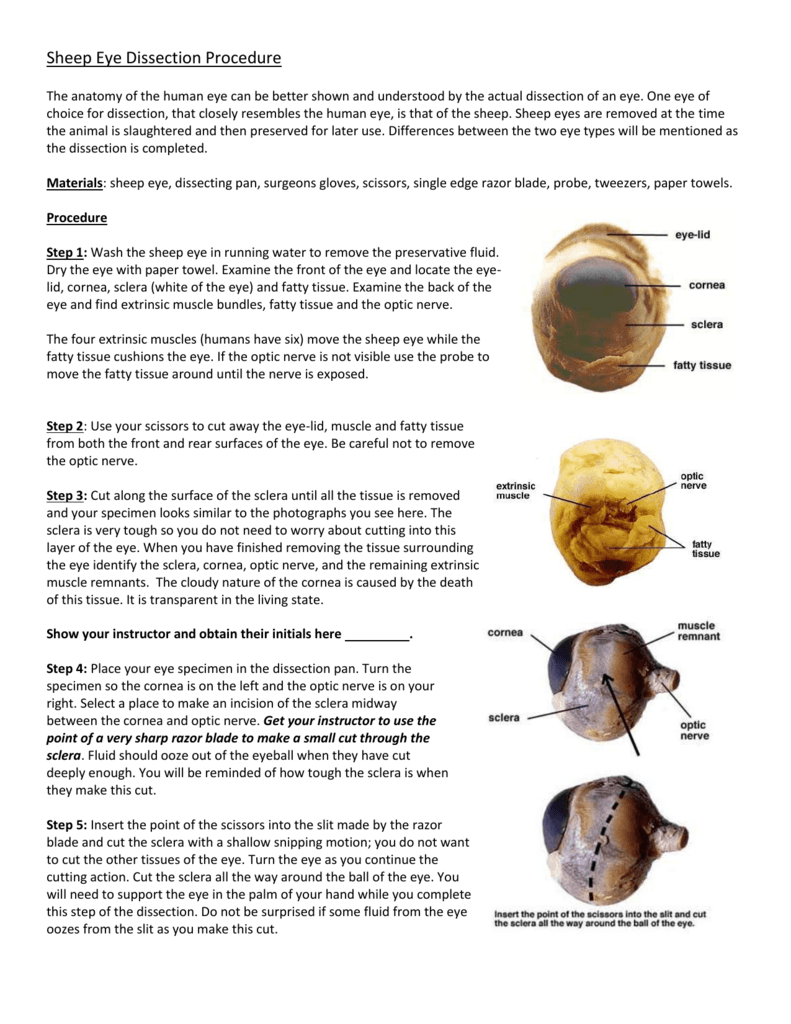
The pineal gland, conarium, or epiphysis cerebri, is a small endocrine gland in the brain of most vertebrates.The pineal gland produces melatonin, a serotonin-derived hormone which modulates sleep patterns in both circadian and seasonal cycles.The shape of the gland resembles a pine cone, which gives it its name. The pineal gland is located in the epithalamus, near the center of the brain ... Sheep Heart Dissection. Sheep have a four-chambered heart, just like humans. By studying the sheep’s anatomy, you can learn how your own heart pumps blood through your body, thereby keeping you alive!. Use this sheep heart dissection guide in a lab for high school students. the white of the eye 80 The intrinsic eye muscles are under the control of which of the following? (Circle the correct response.) autonomic nervous system Dissection of the Cow (Sheep) Eye somatic nervous system 90 What modification of the choroid that is not present in humans is found in the cow eye? COW no What is its function? c 10.
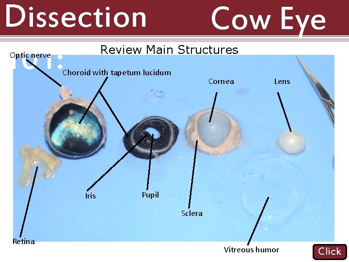
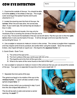
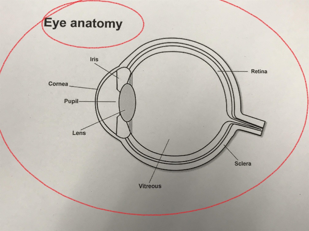
Cow eye dissection diagram. More Dissection Projects: Cow Eye Sheep Heart; Owl Pellet; Frog; Shop other top-selling dissection specimens, such as earthworms for dissection, dogfish shark dissection, dissect crayfish, cow eyes, sheep brain specimen, sheep brain dissection kit, a preserved starfish, or owl pellets for classroom. COW EYE DISSECTION · 1. Examine the outside of the eye. · 2. Locate the covering over the front of the eye, the cornea. · 2. Cut away the fat and muscle, this may ... 1. Place the cow eye on a dissecting tray. The eye most likely has a thick covering of fat and muscle tissue. Carefully cut away the fat and the ... Learn how to dissect a cow's eye in your classroom. This resource includes: a step-by-step, hints and tips, a cow eye primer, and a glossary of terms. Cow's Eye Dissection - Eye diagram
Dissection games Cow eyes are typical dissection specimens used in lab to study eye anatomy because they are structurally and functionally similar to human eyes. thoroughly after the lab. Also, the cow eyes can be ... Place the eyeball on the dissecting tray and gently hold it with two fingers at the cornea and.3 pages Sep 7, 2019 — Step-by-step instructions for a science lab to dissect a cow eye.
Fetal Pig 1 Sheep Brain and Cow Eye Dissection Lab Report Ivy Tech Anatomy and Physiology 101 2/27/2020 Abstract The purpose of the sheep brain and cow eye dissection is to familiarize locating and identify the regions and structures in the brain and eye. Axis vertebra. Pleural cavity 17. The spinal cord is a long, thin, tubular structure made up of nervous tissue, which extends from the medulla oblongata in the brainstem to the lumbar region of the vertebral column.It encloses the central canal of the spinal cord, which contains cerebrospinal fluid.The brain and spinal cord together make up the central nervous system (CNS). In humans, the spinal cord begins at the occipital ... The Cow Eye Dissection Lab. What are the structures of the mammalian eye and how do they function? The mammalian eye consists of many specialized cells and ... the white of the eye 80 The intrinsic eye muscles are under the control of which of the following? (Circle the correct response.) autonomic nervous system Dissection of the Cow (Sheep) Eye somatic nervous system 90 What modification of the choroid that is not present in humans is found in the cow eye? COW no What is its function? c 10.
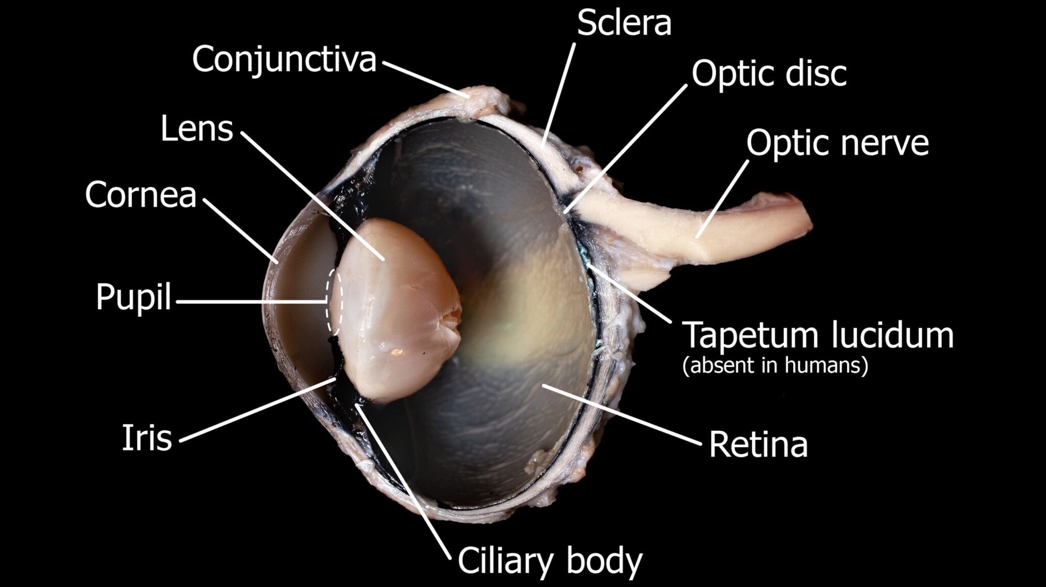
Examine The Outside Of The Eye See How Many Parts Of The Eye You Can Identify You Should Be Able To Find The Whites Or Sclera The Tough Outer Course Hero
Sheep Heart Dissection. Sheep have a four-chambered heart, just like humans. By studying the sheep’s anatomy, you can learn how your own heart pumps blood through your body, thereby keeping you alive!. Use this sheep heart dissection guide in a lab for high school students.

Cow Eye Dissection Perkins Elearning
The pineal gland, conarium, or epiphysis cerebri, is a small endocrine gland in the brain of most vertebrates.The pineal gland produces melatonin, a serotonin-derived hormone which modulates sleep patterns in both circadian and seasonal cycles.The shape of the gland resembles a pine cone, which gives it its name. The pineal gland is located in the epithalamus, near the center of the brain ...

Sheep Eye Dissection Virtual Practical Exam Practice Quiz For Anatomy Youtube

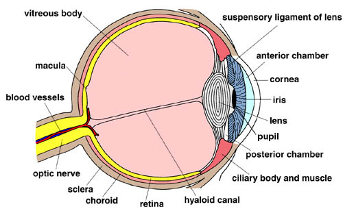
Cow S Eye Dissection Eye Diagram
2

Cows Eyeball Pdf Human Eye Eye

Gttechandinfolit The Different Parts Of The Eye

Elementary Dissection Mat Carolina Cow Eye Carolina Com
2

Cow Eye Dissection Carolina Com
Sheep Eye Dissection Lab

Ppt Sheep Eye Dissection Powerpoint Presentation Free Download Id 2108972
1
Cow Eye Dissection By Tiggerbaby1122 On Deviantart

What Are The Parts Of The Eye Let S Use A Diagram To Help Us Get Familiar With The Parts And Pronounce Them Correctly Diagram Parts Ppt Download

Cow Eye
Click Uplift Education
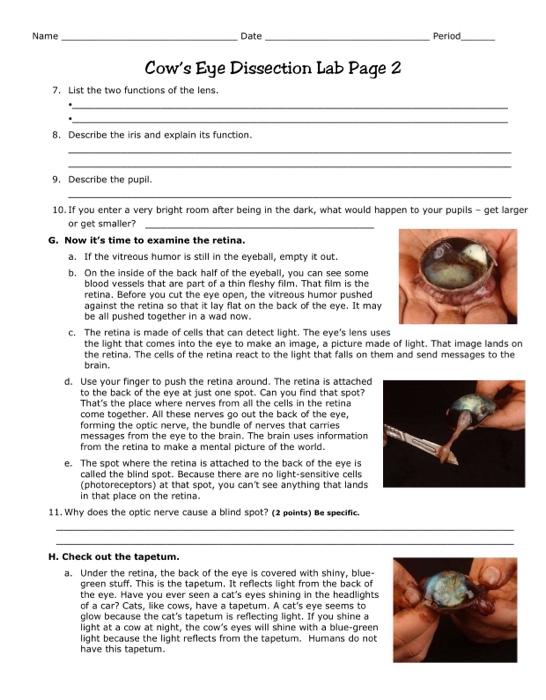
Solved Name Date Period Cow S Eye Dissection Lab Page 2 7 Chegg Com

Cow Eye Dissection Cow Eyes Dissection Diagram Of The Eye
Anatomy Of The Eye Children S Wisconsin
Scb209 Lab3 Natural Sciences Open Educational Resources
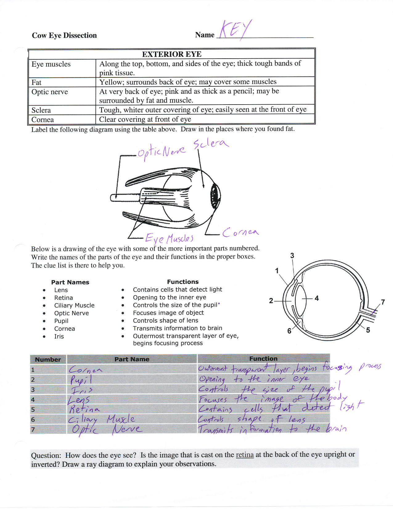
Cow Eye Dissection Key
Dissection 101 Reasons To Use The Dissection Video
2

Cow Eye Dissection Carolina Com
Cow Eye Dissection Anatomy Project Hst Learning Center
Eye Dissection Ppt Download

Wmu Psychology Department Lisa Baker Cow Eyes Dissection How To Memorize Things
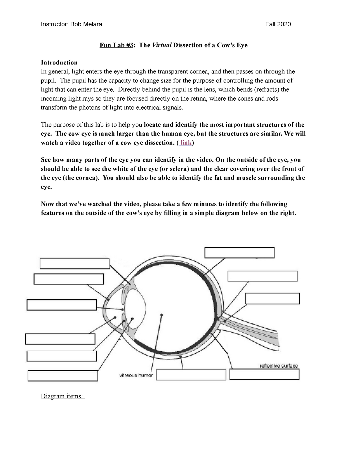
Lab 3 Psy 102 00 1 Instructor Bob Melara Fall 2020 Fun Lab 3 The Virtual Dissection Of A Cow S Studocu
Cow Eye Dissection

Cow Eye Dissection Vocabulary K6 Flashcards Quizlet
Hamburg Csd 5th Grade Cow Eyes
Sheep Eye Dissection Procedures

19 Best Cow Eye Dissection Labeled

Carolina Eye Dissection Mat Carolina Com
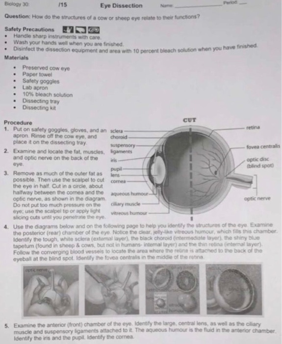
Solved Retina Biology 30 15 Eye Dissection Name Question Chegg Com

Lab 15 Cow Eye Dissection Flashcards Quizlet
2

Mrs Knudson Did Her Famous Cow Eye Vernon Middle School Facebook
0 Response to "39 cow eye dissection diagram"
Post a Comment