35 diagram of muscle fiber
Diagram of the Structure of a Muscle Cell (also called a muscle fibre). The structure of a muscle cell can be explained using a diagram labelling muscle filaments, myofibrils, sarcoplasm, cell nuclei (nuclei is the plural word for the singular nucleus), sarcolemma, and the fascicle of which the muscle fibre is part. The structure of muscle fibers is included in courses in human biology and ...
FG fibers are used to produce rapid, forceful contractions to make quick, powerful movements. These fibers fatigue quickly, permitting them to only be used for short periods. Most muscles possess a mixture of each fiber type. The predominant fiber type in a muscle is determined by the primary function of the muscle.
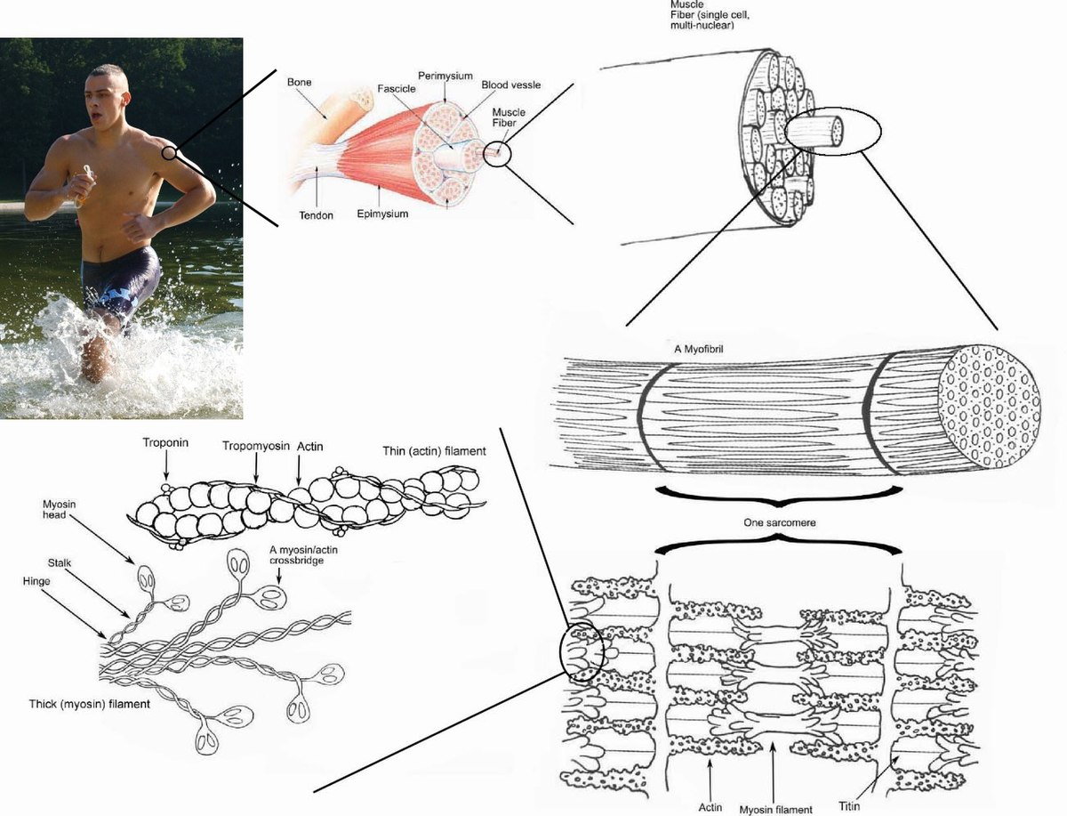
We'll just keep zooming until we get to the molecular level. So this myofibril, which is-- remember, it's inside of the muscle cell, inside of the myofiber. The myofiber is a muscle cell. Myofibral is a-- you can view it as a tube inside of the muscle cell. These are the things that are actually doing the contraction.

Diagram of muscle fiber
Skeletal Muscle Fiber Location and Arrangement. are located inside muscles, where they are organized into bundles called […] Internal Anatomy of Skeletal Muscle Fibers. An interactive quiz about the internal anatomy of skeletal muscle fibers, featuring illustrations-based multiple choice questions. Skeletal Muscle Fiber Location and Arrangement > Internal Anatomy of Skeletal Muscle Fibers ...
Blank Muscle Diagram Unlabeled : Muscle Fiber Diagram Unlabeled Png Image Transparent Png Free Download On Seekpng : Posted by Larry Napier on Kamis, 11 November 2021 In this chapter we describe the gross anatomy of the muscular system and consider functional relationships between muscles and bones of the body .
Start studying Anatomy: Muscle Fiber Diagrams. Learn vocabulary, terms, and more with flashcards, games, and other study tools.
Diagram of muscle fiber.
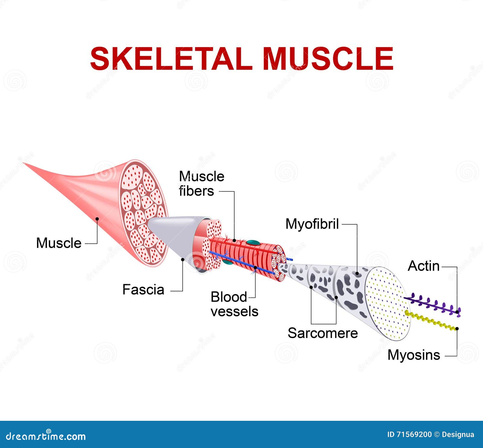
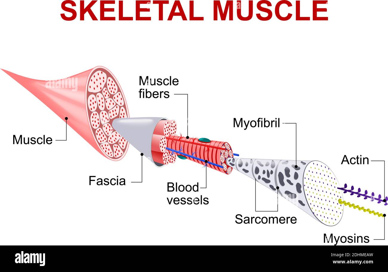
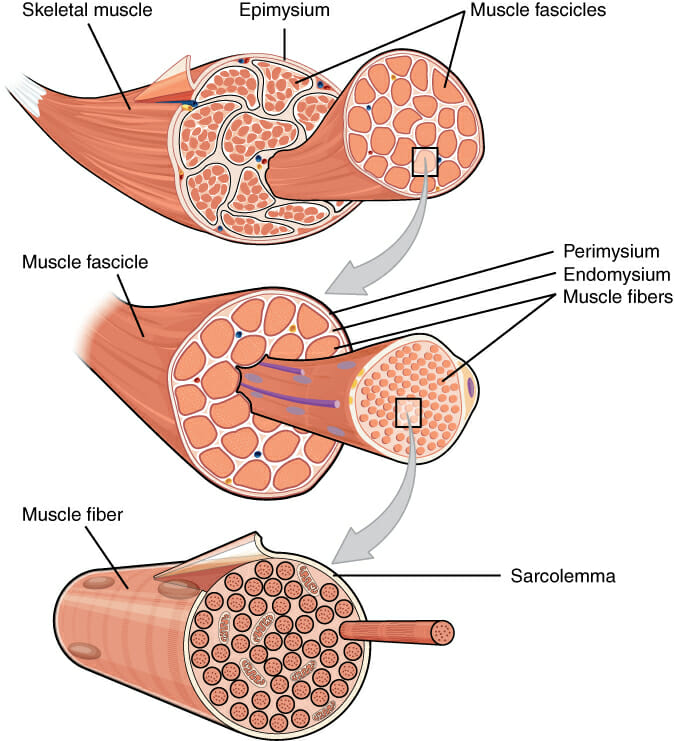
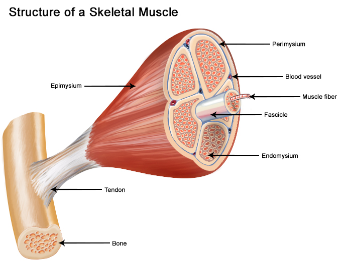
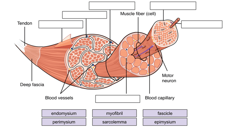
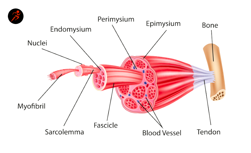
Each compartment contains a bundle of muscle fibers. Each bundle of muscle fiber is called a fasciculus and is surrounded by a layer of connective tissue called ...
Label this diagram of a muscle fiber, using these terms: myofibril, Z line, T tubule, sarcomere, sarcolemma, sarcoplasmic reticulum. Step-by-step solution Chapter 39, Problem 19TY is solved.
bundle and a single axon may innervate a few to 100's of muscle fibers at same time each muscle organ is composed of 1000's of motor units whole motor unit responds as "all or none" muscle cells cannot "partially" contract the fewer muscle cells/ motor unit more precise movement the muscle can make eg. eye: 10-23 fibers/axon hand: few
The musculoskeletal system comprises one of the major tissue/organ systems in the body. The three main types of muscle tissue are skeletal, cardiac, and smooth muscle groups.[1][2][3] Skeletal muscle attaches to the bone by tendons, and together they produce all the movements of the body. The skeletal muscle fibers are crossed with a regular pattern of fine red and white lines, giving the ...
Muscle Fiber Diagram Unlabeled is a high-resolution transparent PNG image. It is a very clean transparent background image and its resolution is 1210x849 , please mark the image source when quoting it.
Muscle Charts of the Human Body For your reference value these charts show the major superficial and deep muscles of the human body. Superficial and deep anterior muscles of upper body
12+ Cardiac Muscle Diagram. Cardiac muscle tissue is made up of many interlocking cardiac muscle cells, or fibers, that give the tissue its properties. This is one feature that differentiates it from skeletal. Contraction of Cardiac Muscle - Pathway of Contraction … from teachmephysiology.com
Muscle Fibres · Skeletal muscles consist of tightly packaged muscular bundles (fascicles) surrounded by connective tissue ( · Each bundle contains multiple muscle ...
Figure 9.13b A motor unit consists of a motor neuron and all the muscle fibers it innervates. Branching axon to motor unit Muscle fibers Axon terminals at neuromuscular junctions (b) Branching axon terminals form neuromuscular junctions, one per muscle fiber (photomicrograph 330x).
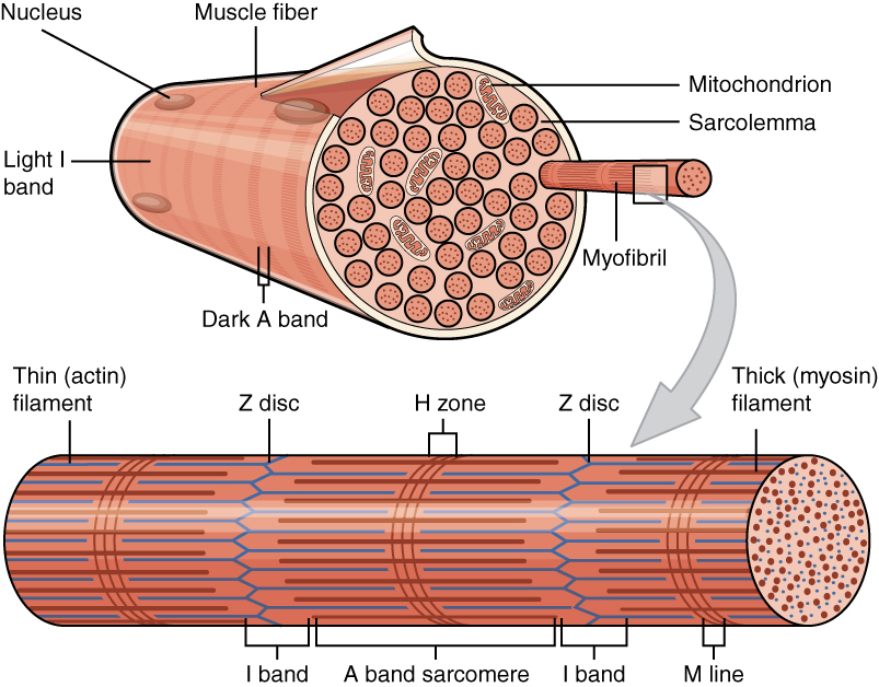
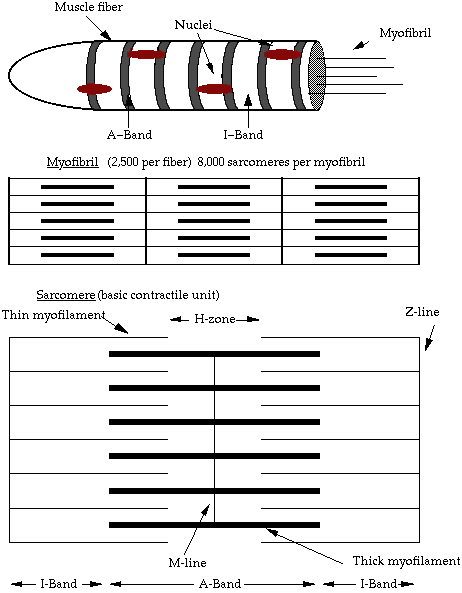
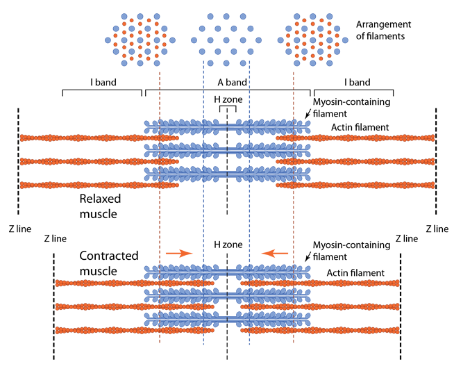
Diagram of part of a muscle fiber showing the myofibrils. One myofibril extends from the cut end of the fiber. Small part of one myofibril enlarged to show the myofilaments responsible for the banding pattern. Each sarcomere extends from one Z disc to the next. Enlargement of one sarcomere (sectioned lengthwise).
Muscle Fiber Diagram Unlabeled. 1210*849. 0. 0. PNG. Images For Ear Anatomy Diagram Blank - Unlabelled Diagram Of The Ear. 1280*975. 0. 0. PNG. 6th Grade Muscle Diagram. 591*684. 0. 0. PNG. Small - Chloroplast Diagram Unlabeled. 516*596. 0. 0. PNG. The Peripheral Nervous System Is The Part Of The Nervous - Nervous System Diagram Unlabeled.
Muscle fibers are classified on the basis of their physiological behavior as fast (or fatigable) or slow (fatigue resistant), largely an expression of aerobic enzyme activity and calcium metabolism, and by myosin heavy chain isoform into type 1, types 2A, 2B and 2 ×. Type 2B is not expressed in human skeletal muscle. With only a few exceptions, each motor unit is composed of fibers of one ...
Muscle fiber diagram - shameless pride
In the diagram, individual muscle fibers are covered by this layer. a) K b) I c) L d) M e) A. c. In the diagram, one of the components of a myofibril is the structure labeled _____.? a) G b) H c) B d) J e) None of these choices are correct. b. In the diagram, what is the basic functional unit of a myofibril? a) B

10.2 skeletal muscle – anatomy & physiology
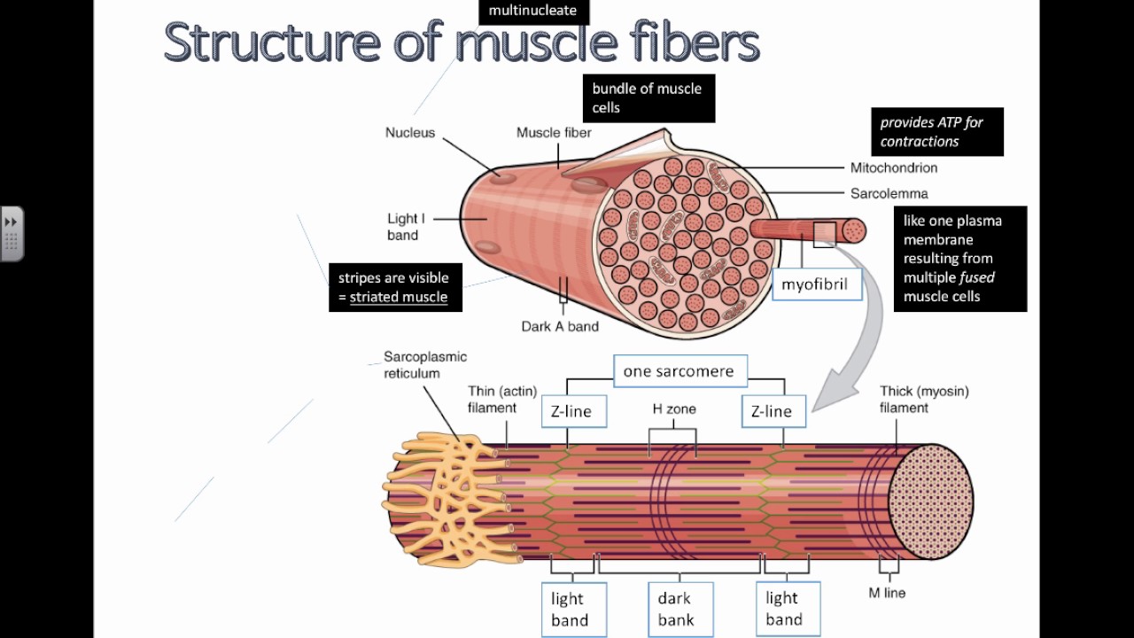
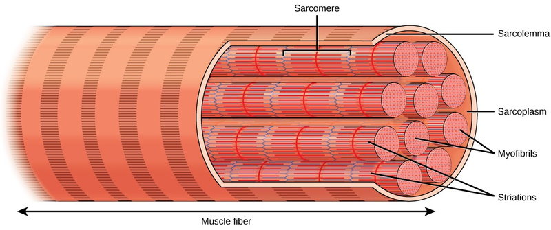
Each skeletal muscle fiber is a skeletal muscle cell. Within each muscle fiber are myofibrils, long cylindrical structures that lie parallel to the muscle fiber.Myofibrils run the entire length of the muscle fiber. They attach to the plasma membrane, called the sarcolemma, at their ends, so that as myofibrils shorten, the entire muscle cell contracts (Figure 16.18).
Muscle fiber stock illustrations – 3,373 muscle fiber stock ...
Muscle fibers are single muscle cells. When grouped together, they work to generate movement of your body and internal organs. You have three types of muscle tissue: skeletal, smooth, and cardiac.

Muscles of the body exposed | skeletal muscle, muscle diagram ...
Start studying Anatomy: Muscle Fiber Diagrams. Learn vocabulary, terms, and more with flashcards, games, and other study tools.

Muscle-skeletal muscle-gross and ultra structure - online biology ...
Each muscle is comprised of thousands of muscle fibers that are bundled together. Skeletal muscle is predominantly involved in movement. When one of these muscles contracts, it allows movement of a specific area of the body. Smooth muscles. Smooth muscle can be found in many different organ systems of your body, including but not limited to your digestive system, respiratory system ...
Structure of muscle fibers (ib biology)
The human body has three different types of muscles. They include: skeletal muscles, smooth muscles, cardiac muscles. Skeletal muscles. Your skeletal muscles are attached to your bones via tendons. Each muscle is comprised of thousands of muscle fibers that are bundled together. Skeletal muscle is predominantly involved in movement.
Skeletal muscle fiber high resolution stock photography and images ...
Labels/Diagram based on Figure 6.3 Anatomy of Skeletal Muscle Fiber
Skeletal muscle fiber structure and function | open textbooks for ...
A muscle cell is very specialised for its purpose. One muscle cell is known as a muscle fibre, and its cell surface membrane is known as the sarcolemma.

Sarcoplasm - an overview | sciencedirect topics
The primary metabolic pathway used by a muscle fiber determines whether the fiber is classified as oxidative or glycolytic. If a fiber primarily produces ATP through aerobic pathways it is oxidative. More ATP can be produced during each metabolic cycle, making the fiber more resistant to fatigue. Glycolytic fibers primarily create ATP through anaerobic glycolysis, which produces less ATP per ...
File:1002 organization of muscle fiber.jpg - wikimedia commons
muscle cell or muscle fiber ↓ fascicles ↓ whole skeletal muscle Page 11. Pyramid of Subunits • Whole muscle as a pyramid of subunits: Fascicles Muscle cells (Muscle Fibers) Myofibrils Myofilaments Page 12. Summary • The three types of muscle cells in the body are skeletal, cardiac, and smooth.

Histology of muscle | muscle anatomy, skeletal muscle anatomy ...
Figure 10.2.2 - Muscle Fiber: A skeletal muscle fiber is surrounded by a plasma membrane called the sarcolemma, which contains sarcoplasm, the cytoplasm of muscle cells. A muscle fiber is composed of many myofibrils, which contain sarcomeres with light and dark regions that give the cell its striated appearance.

Skeletal muscle fiber labeled
Skeletal muscle is made up of thousands of muscle fibres that run the length of the muscle. Each muscle fibre consists of many contractile units called myofibrils which run the length of each muscle fibre. Individual muscle fibres are wrapped with fascia and then further bound together by more fascia into bundles called fascicules.

Muscle fiber anatomy in detail
Diagram of different stem cells Diagram of different stem cells illustration muscle fiber stock illustrations Diagram of different stem cells Motor neuron Motor neurone is a cell body is located in the brain, or the spinal cord.

The structure of a skeletal muscle fiber | biology lessons, human ...
Relaxing skeletal muscle fibers, and ultimately, the skeletal muscle, begins with the motor neuron, which stops releasing its chemical signal, ACh, into the synapse at the NMJ. The muscle fiber will repolarize, which closes the gates in the SR where Ca ++ was being released. ATP-driven pumps will move Ca ++ out of the sarcoplasm back into the SR.

Muscle fiber diagram unlabeled png image | transparent png free ...
FG fibers are used to produce rapid, forceful contractions to make quick, powerful movements. These fibers fatigue quickly, permitting them to only be used for short periods. Most muscles possess a mixture of each fiber type. The predominant fiber type in a muscle is determined by the primary function of the muscle.

Ultrastructure of muscle - skeletal - sliding filament ...
Structure of muscle fibre showing a sarcomere under electron microscope with schematic explanation. Diagram of sarcoplasmic reticulum with terminal cisternae ...
Muscle - definition, function, types and structure | biology ...
Muscle Fiber Diagram.Diagram with myofibril and muscle fibers. Diagram of the Structure of a Muscle Cell (also called a muscle fibre). PT on the Net (Esther Aguilar) Diagram with myofibril and muscle fibers. There is a printable worksheet available for download here so you can take.

Muscle fiber diagram | quizlet
Skeletal muscle is made up of thousands of muscle fibres that run the length of the muscle. Each muscle fibre consists of many contractile units called myofibrils which run the length of each muscle fibre. Individual muscle fibres are wrapped with fascia and then further bound together by more fascia into bundles called fascicules.

Skeletal muscle fiber diagram | quizlet
Muscle cramps occur when a single skeletal muscle fiber, muscle, or entire muscle group contracts involuntarily. They’re often painful and can last for several seconds or minutes.

Label the following in a diagram of a skeletal muscle fiber ...
The fast muscle (what the researchers call type IIa) moves 5 times faster than the slow muscle, and the super-fast (called type IIb) moves 10 times faster than the slow muscle fiber. The average person has approximately 60% fast muscle fiber and 40% slow-twitch fiber (type I). There can be swings in fiber composition, but essentially, we all ...
Seer training: structure of skeletal muscle
Each skeletal muscle fiber is a skeletal muscle cell. These cells are incredibly large, with diameters of up to 100 µm and lengths of up to 30 cm.
Muscles labeling
SKELETAL MUSCLE FIBER DIAGRAM. Image: Skeletal Muscle Fibers. When viewing the I bands within the myofibril, it can be clearly seen that I bands are considered to run from one edge of the stack of filaments to the next. They take on a lighter shade due to the thin fibers of their creation. Despite the appearance of a distinctive border, the I ...

Diagrammatically show the difference between the three types of ...
Diagram the process of cross-bridge cycling; The Neuromuscular Junction. The process of muscle contraction begins at the site where a motor neuron's terminal meets the muscle fiber—called the neuromuscular junction (NMJ). Every skeletal muscle fiber in every skeletal muscle is innervated by a motor neuron at a NMJ. Excitation signals from ...
![3: Structure of a muscle fiber. Image: [Spe98] | Download ...](https://www.researchgate.net/profile/Kolja-Kaehler/publication/47861059/figure/fig2/AS:669428580642827@1536615619762/Structure-of-a-muscle-fiber-Image-Spe98.png)
3: structure of a muscle fiber. image: [spe98] | download ...
Structure and composition of muscle - meat science

Diagram showing the level of organization within a skeletal muscle ...
Skeletal muscle - wikipedia

Muscle fiber anatomy quiz

Skeletal muscle physiology

Skeletal muscle | anatomy and physiology i
Muscle fibers explained: type i and type ii (slow & fast twitch ...
The muscular system

Skeletal muscle diagram the muscular system micro and macro anatomy

The structure of skeletal muscle. striated muscle fiber consists ...
0 Response to "35 diagram of muscle fiber"
Post a Comment