37 pressure ulcer sites diagram
When selecting codes for location of pressure ulcers involving the posterior lower ... or the trochanters, all of which are common sites for pressure ulcer ... A pressure ulcer is an ulcer related to some form of pressure and should not be confused with ulcers relating to disease (like cancer), vascular flow (venous or arterial) or neuropathy (like in persons with diabetes) You should be able to see a “cause and effect” relating to pressure with the ulcer.
by K Agrawal · 2012 · Cited by 213 — Pressure ulcer in an otherwise sick patient is a matter of concern for the care ... the contribution of Ms. Shrreya Agrawal for preparing the line diagram.

Pressure ulcer sites diagram
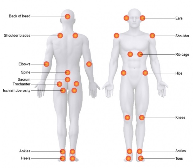
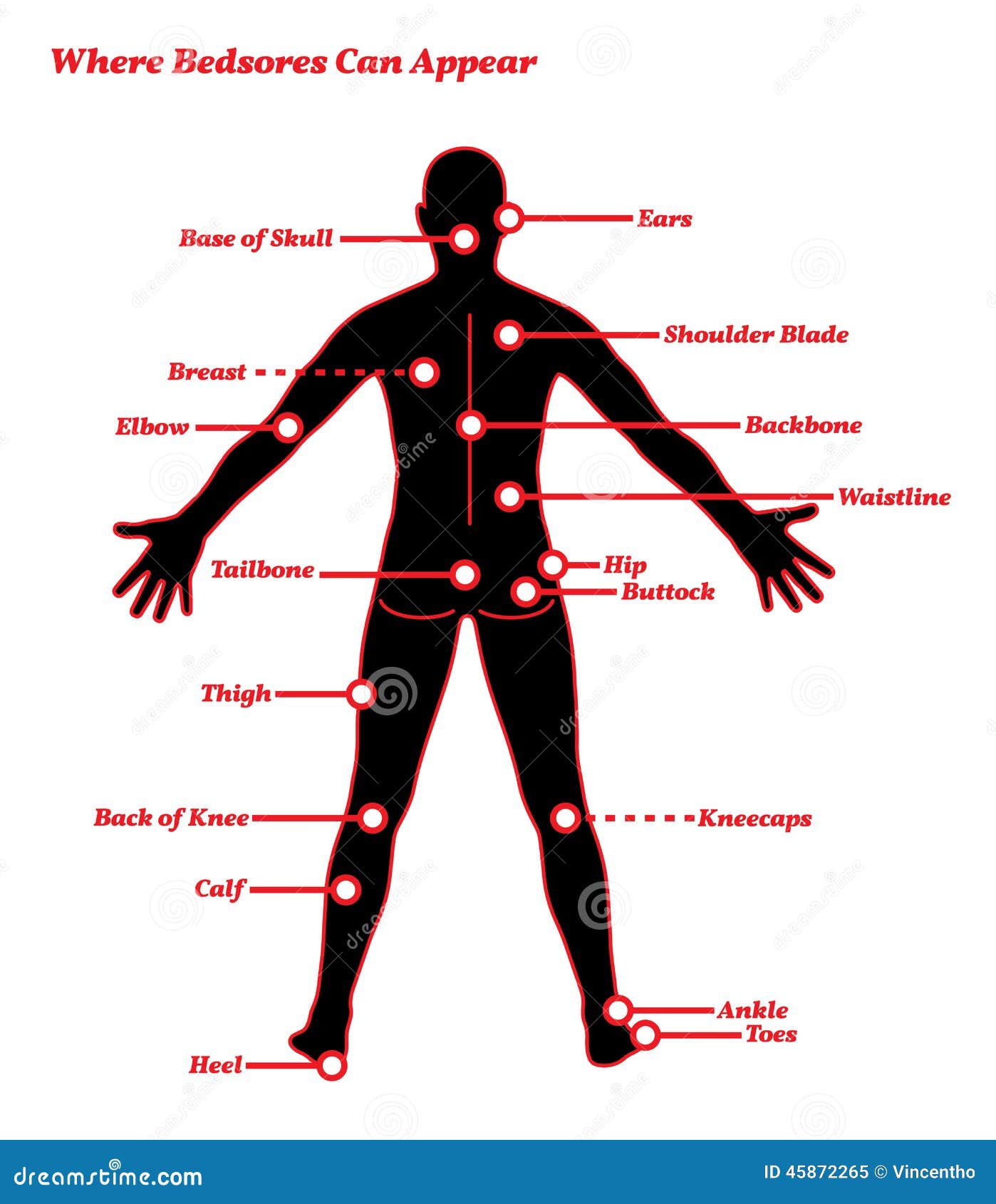
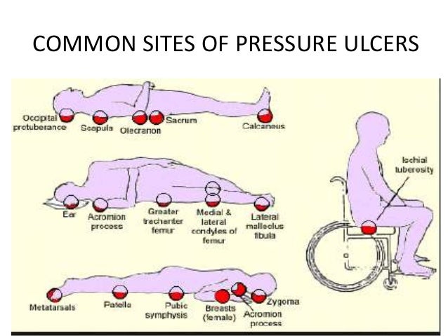
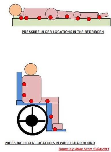
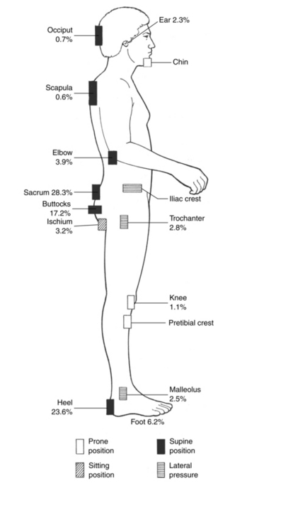
Skeleton presentation–Common sites of pressure ulcers. The most common sites are the sacrum, greater trochanters, ischial tuberosities, medical and lateral ... Find out more about the causes of pressure sores and how to prevent them. ... Diagram showing the areas of the body at risk of pressure sores when sitting ... The most common sites are the back of the head and ears, the shoulders, the elbows, the lower back and buttocks, the hips, the inner knees, and the heels.Missing: diagram | Must include: diagram
Pressure ulcer sites diagram. What Is a Pressure Ulcer? Pressure ulcers are wounds caused by the pressure of body weight on your skin covering bony areas.1 Pressure ulcers first appear ...Missing: diagram | Must include: diagram The depth of a Category/Stage III pressure ulcer varies by anatomical location. ... In contrast, areas of significant adiposity can develop extremely deep ... Common sites of pressure ulcers when sitting include the shoulder blade, the buttocks, the. Sitting. When lying on the side, pressure ulcers can occur on ... Pressure ulcer risk assessment is a standardized and ongoing process with the goal of identifying patients at risk for the development of a pressure ulcer so that plans for targeted preventive care to address the identified risk can be implemented. This process is multifaceted and includes many components, one of which is a validated risk ...
Areas where bones are close to the surface (called "bony prominences") and areas that are under the most pressure are at greatest risk for developing ... A professional is providing skin care (treatment) for a client who has a pressure ulcer. The HCS/AAA/DDD Social Worker/Nurse or other contracted nursing resource must: Verify . with [Health Care Provider Name (HCP)] by ( ) that a treatment plan is in place for the client’s pressure ulcer(s). Jan 31, 2019 · Pressure ulcer risk assessment using clinical judgement alone: Braden pressure ulcer risk assessment and training: Pressure ulcer incidence Visual skin assessment Follow‐up: 8 weeks: Study population: RR 1.43 (0.77 to 2.68) 65 more per 1000 (from 35 fewer to 254 more) 180 (1 study) ⊕⊝⊝⊝ Very low 1 Feb 01, 2018 · Top diagram showing pressure ulcers Stage I: skin intact. Stage II: partial skin loss. Stage III: full-thickness skin loss, subcutaneous tissue exposed. Stage IV: muscle, tendon, bone or organs exposed. Bottom diagram showing unstageable pressure ulcer with tissue damage hidden from observer by eschar over entire wound. Deep tissue injury ...
Secondary hypertension is hypertension due to an identifiable cause, and may result in certain specific additional signs and symptoms. For example, as well as causing high blood pressure, Cushing's syndrome frequently causes truncal obesity, glucose intolerance, moon face, a hump of fat behind the neck and shoulders (referred to as a buffalo hump), and purple abdominal stretch marks. –Pressure ulcer –Rash –Infection, cellulitis •Deficiencies can also affect skin: –Vitamin C deficiency causes purplish blotches on lightly traumatized areas. –Zinc deficiency causes redness of the nasolabial fold and eyebrows. 17 The most common sites are the back of the head and ears, the shoulders, the elbows, the lower back and buttocks, the hips, the inner knees, and the heels.Missing: diagram | Must include: diagram Find out more about the causes of pressure sores and how to prevent them. ... Diagram showing the areas of the body at risk of pressure sores when sitting ...
Skeleton presentation–Common sites of pressure ulcers. The most common sites are the sacrum, greater trochanters, ischial tuberosities, medical and lateral ...











0 Response to "37 pressure ulcer sites diagram"
Post a Comment