40 where on the diagram is the femoral area?
In the diagram,the femoral area is _____to the cervical area? 11ea7f55_b8e6_2760_9ecd_a1ab91efd6f7_TB6091_00 A)superior B)inferior C)medial D)proximal E)posterior Ask a new question Ask a question
Hip Anatomy, Function and Common Problems. June 29, 2021. July 28, 2010 by Dr. Andrew Chung. The hip joint is a ball-and-socket type joint and is formed where the thigh bone (femur) meets the pelvis. The femur has a ball-shaped head on its end that fits into a socket formed in the pelvis, called the acetabulum.
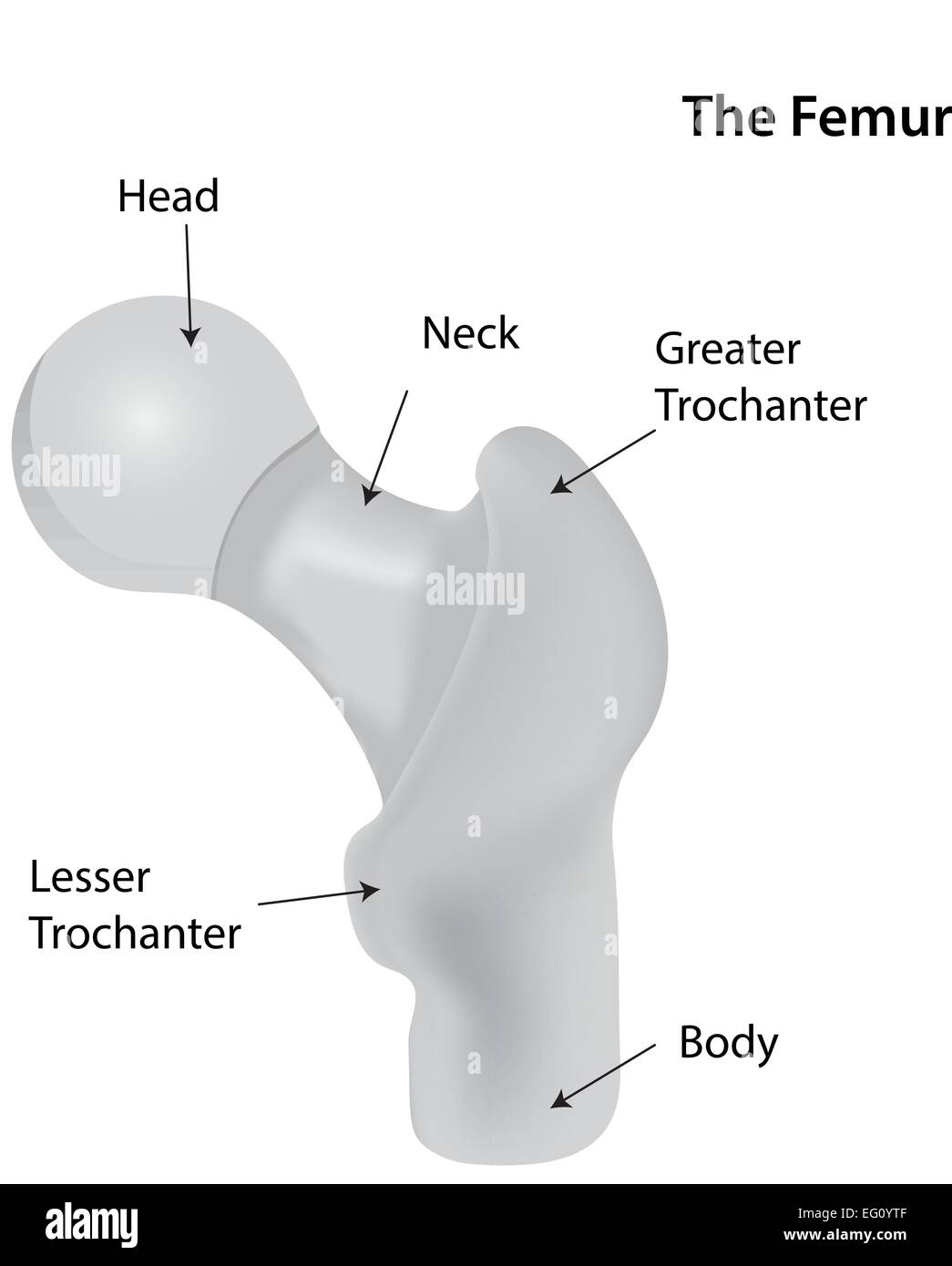
The femur, or thigh bone, is the proximal bone of the hindlimb in tetrapod vertebrates. ... The transition area between the head and neck is quite rough due to ...

Where on the diagram is the femoral area?
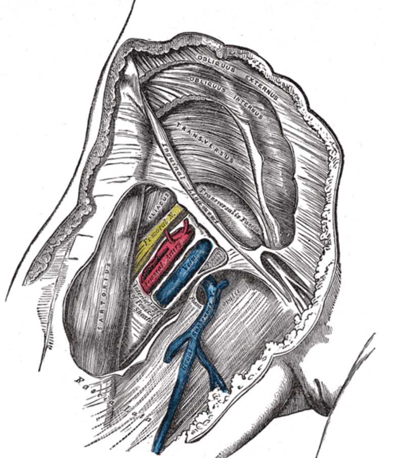
This photo about: Where On the Diagram is the Femoral area, entitled as Superficial Vessels And Nerves Of The Anterior Trunk Pectoral Where On The Diagram Is The Femoral Area - also describes Superficial Vessels and Nerves of the Anterior Trunk Pectoral and labeled as: where in my ip,where in queens is the king of queens,where in san go,where in zanzibar to stay,where to visit, with resolution ...
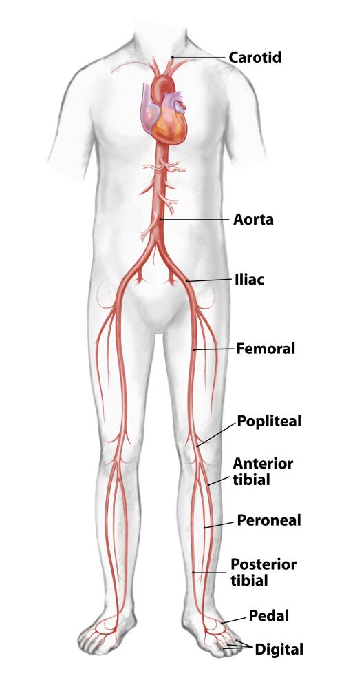
We are pleased to provide you with the picture named Human Body Artery Diagram In Detail.We hope this picture Human Body Artery Diagram In Detail can help you study and research. for more anatomy content please follow us and visit our website: www.anatomynote.com. Anatomynote.com found Human Body Artery Diagram In Detail from plenty of anatomical pictures on the internet.
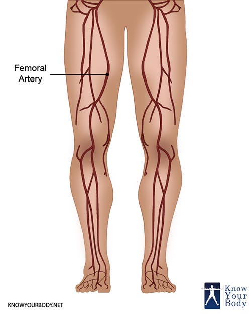
13 Femoral Artery Pain. The Femoral Artery is a term used for a group of few arteries which passes fairly close to the outer surface of the thighs. It begins at the inguinal ligament, called the Femoral Head, and ends just above the knee at p place called the Adductor canal or the hunter's canal. It divides into smaller branches so as to ...
Where on the diagram is the femoral area?.
Hey all, I'm a slim, 21-year-old male. I have a rabbit-hole of doctor visits/scans that I've done in the last 6 months, but none have produced even a remotely accurate diagnosis, let alone a diagnosis at all. Let me give you a quick rundown/timeline: **July 2018:** Had right femoral hernia surgery after what was most-likely a work-out injury. Took about a month to fully recover from this and get back into my daily routine. **\~ October 2018:** I began having a very infrequent twitching on the ...
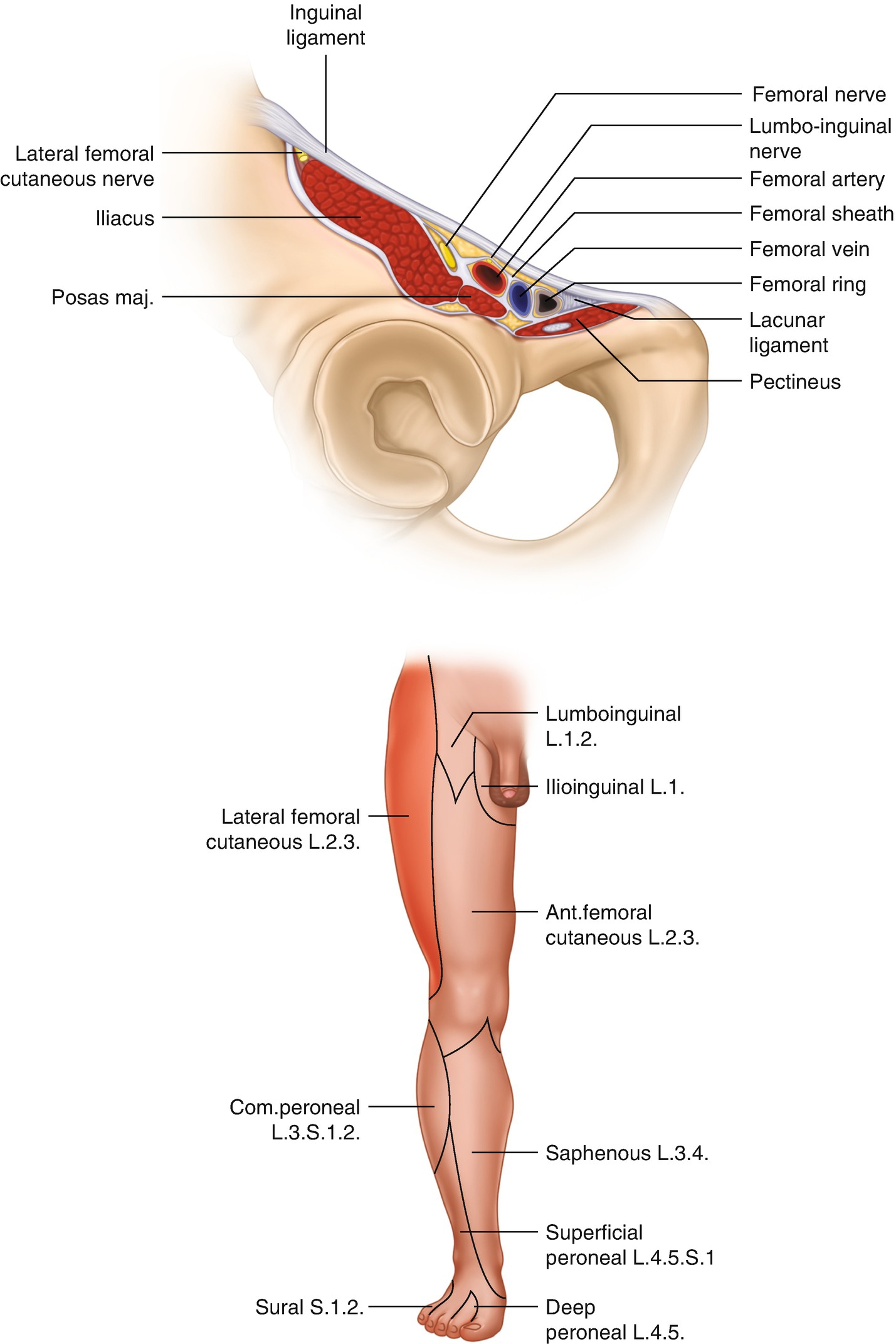
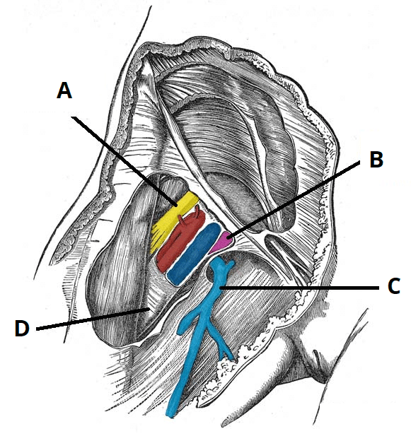
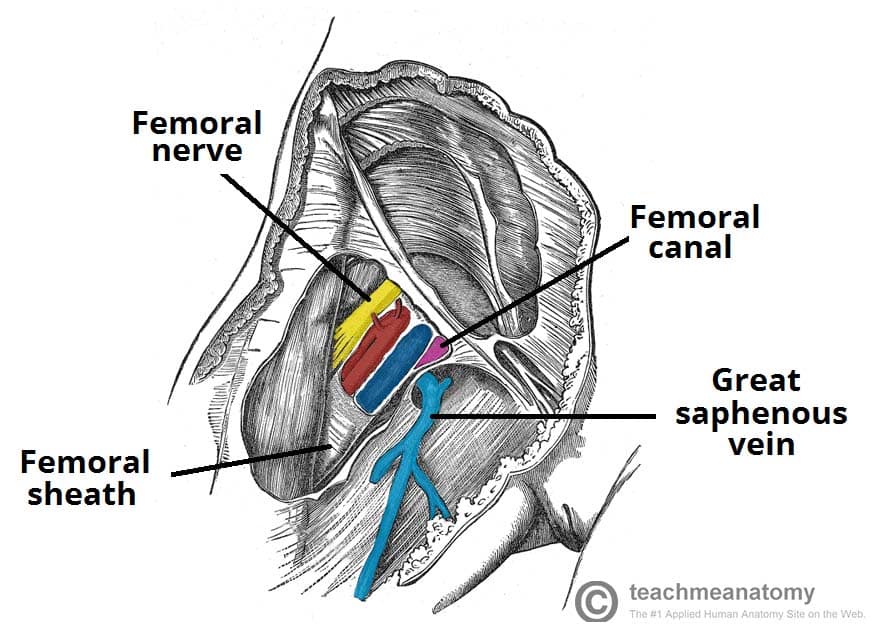
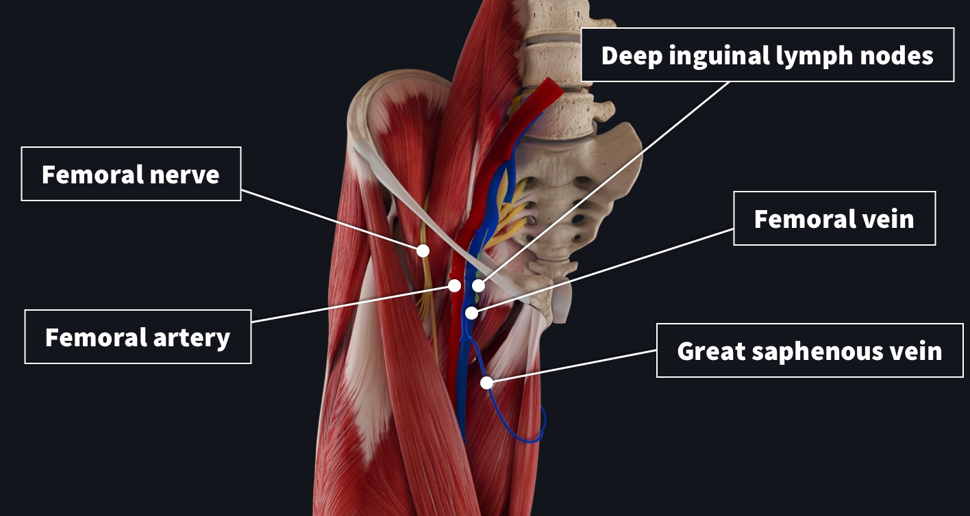
The femoral triangle is a hollow area in the thigh through which many large nerve structures pass. It is an important area as it has both clinical and anatomical significance. The femoral artery is enclosed within the femoral sheath along with the femoral vein. Femoral Artery Location .
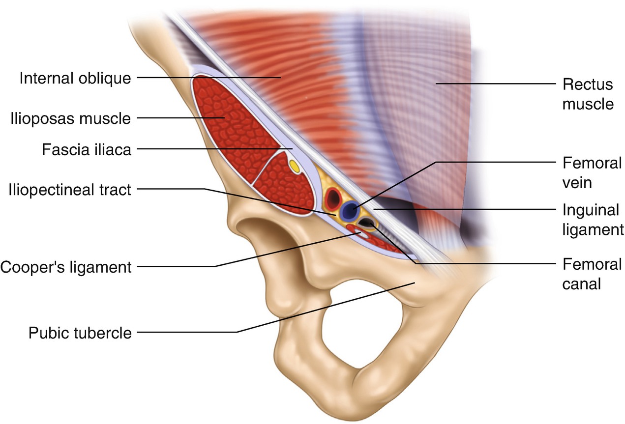
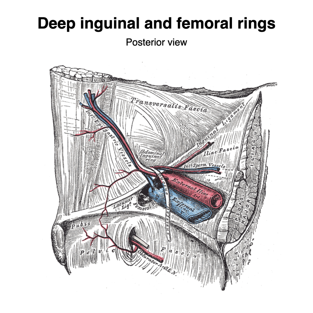
Femoral Hernia: Femoral hernia is a type of hernia when weak tissues in the groin or inner thigh push through a weak spot. It causes severe discomfort in the groin and is not common. To treat femoral hernia, surgery becomes necessary as it may result in complications. Femoral hernia can occur from straining or excess pressure in the groin.
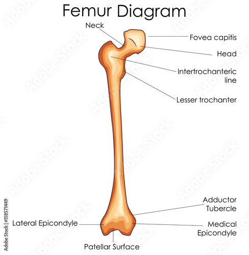
Femur. The femur is the only bone located within the human thigh. It is both the longest and the strongest bone in the human body, extending from the hip to the knee. Important features of this ...
Femoral (superficial inguinal) C774 Inguinal region or leg Inguino-femoral, right and left* Foramen of Winslow (epicolic, omental) C772 Intra-abdominal Mesenteric Gastric (inferior, left, right, superior, NOS) C772 Intra-abdominal Mesenteric September 2021 Summary Stage 2018 Coding Manual v2.1 2
Unit 1 Picture questions. Where on the diagram is the femoral area? B) They act as the receptors. Study the diagram. What is the role of the stretch-sensitive nerve cells in the cervix? A) They act as the stimulus. B) They act as the receptors.
The femoral triangle is a wedge-shaped area located within the superomedial aspect of the anterior thigh.. It acts as a conduit for structures entering and leaving the anterior thigh.. In this article, we shall look at the anatomy of the femoral triangle - its borders, contents, and clinical relevance.
Common femoral artery: This first part of the femoral artery is an extension of the external iliac artery in the pelvis. It contains several branches that supply blood to the tissues in the abdominal wall, groin and pubic area. Deep femoral artery: This artery branches off the common femoral artery. It supplies blood to the femur, hip, buttocks ...
View Test Prep - SC121 Quiz 1 from SC 121 at Kaplan University. SC121 Quiz 1 1. Where on the diagram is the femoral area? Thigh area 2. What is the name of the outer layer of the serous membrane that
Femur, upper bone of the leg or hind leg. The head forms a ball-and-socket joint with the hip (at the acetabulum), being held in place by a ligament within ...
The femoral nerve is the major nerve that serves the tissues of the thigh and leg, including the muscles and skin. While the much larger sciatic nerve also passes through the thigh on its way to the lower leg and foot, only the femoral nerve innervates the tissues of the thigh.
The bones of the hip include the femur, the ilium, the ischium, and the pubis. The pubis, ischium, and ilium together constitute the pelvis while the thigh bone is the femur. The bones together make up the hip. The hip itself is a ball and socket joint, much like the shoulder.The structures necessary to create this joint are the socket, the joint capsule, muscle, ligaments, and the neck ...
Zones and branches of deep femoral artery | download ...
The femoral triangle is important as a number of vital structures pass through it, right under the skin. The following structures are contained within the femoral triangle (from lateral to medial): . Lateral cutaneous nerve of thigh - It crosses the lateral angle of the triangle, runs on the lateral side of the thigh and ends by dividing into anterior and posterior branches.

Femoral triangle | pdf | lower limb anatomy | human anatomy
Femoral nerve (Nervus femoralis) The femoral nerve is a mixed nerve of the lower limb that innervates the muscles and skin of the hip and thigh.. The femoral nerve originates from the lumbar plexus, arising from the anterior rami of spinal nerves L2-L4. In fact, it is the longest branch of the lumbar plexus. The femoral nerve is split by the lateral circumflex femoral artery into an ...
Femur labeled diagram stock vector image & art - alamy
In the diagram the femoral area is to the cervical. 26) In the diagram, the femoral area is _____to the cervical area? a) superiorb) inferior c) mediald) proximal e) posterior Answer: b.
Anatomy of the femoral region | springerlink
**Click this link to go straight to the photographic timeline and 4K word analysis of this faked event**: [False Flag Theater: Boston Bombing Involves Clearly Staged Carnage](http://truthandshadows.com/2013/05/08/false-flag-theatre-boston-bombing-involves-clearly-staged-carnage/) [Paul Craig Robert's endorsement of my analysis from 4/15/17](http://www.paulcraigroberts.org/2017/04/15/boston-marathon-bombing-four-years/) Dr. Paul Craig Roberts has had careers in scholarship and academia, journ...
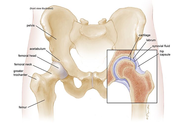
Anatomy of the hip - mu health care
Femur bone anatomy made easy using a labeled diagram of the main parts ... and blood vessels including the popliteal artery, popliteal vein, ...Mar 29, 2021 · Uploaded by EZmed

Femoral artery - wikipedia
Anatomy of the groin area superficial muscles and deep muscles. In this image, you will find rectus abdominis, external oblique, inguinal ligament, tensor fascia lata, gracilis, sartorius, rectus femoris, the iliotibial band in it. You may also find transversus abdominis, iliopsoas, gluteus medius, pectineus, adductor longus, adductor brevis ...
Anatomy of the femoral region | springerlink
The femoral artery is one of the major arteries in the human body. It extends from the iliac artery near the abdomen down to the legs. The primary function of this artery is to supply blood to the ...

Femoral artery - location, anatomy, branches, function and ...
Download scientific diagram | Computerized tomography image, schematic diagram, and nerve conduction study results. A computerized tomography image of the femoral artery and vein area, 2 weeks ...

Femur - wikipedia
Human Anatomy Diagram - This Diagrams Shows The Major Arteries In The Human. deep femoral vein anatomy function & diagram the femoral vein is a vein running alongside the femoral artery the femoral artery is located in the upper area of the thigh and consists of multiple arteries the deep femoral vein also known as the profunda femoris vein ...
Medical education chart of biology for femur bone diagram ...
The femoral triangle is a hollow region located in the supero-medial part of the anterior thigh. It appears most prominently with hip flexion, abduction and internal rotation. It is an easily accessible area through which multiple neurovascular structures pass through. This anatomical landmark is mostly used in dissection and describing ...

Anatomy of femoral area showing close proximity of vein ...
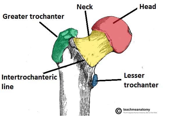
The femoral neck is about 5 cm long and can be subdivided into three regions.The most lateral aspect (the part closest to the greater trochanter) is known as the base of the femoral neck or the basicervical portion of the neck is the widest part of the neck of the femur.The middle segment is also referred to as the midcervical part and is the narrowest part of the femoral neck.
Femoral region
Where on the diagram is the femoral area a D b E c F d J e K Answer a Difficulty from ANATOMY 208 at Queensborough Community College, CUNY.
The femoral triangle - borders - contents - teachmeanatomy
The femoral artery ( FA) is the continuation of the external iliac artery (EIA) at the level of the inguinal ligament. As well as supplying oxygenated blood to the lower limb, it gives off smaller branches to the anterior abdominal wall and superficial pelvis. On this page:
The femoral triangle - borders - contents - teachmeanatomy
femoral vein, which is located medial to the common femoral artery. The inguinal ligament is the landmark that divides the common femoral vein from the external iliac vein (Fig. 3). Approach It is convenient for the US examination to begin with the patient in a supine or semi-Fowler position. The reverse Trendelenburg position,
Vascular anatomy | lemaitre
A diagram of the knee, showing the femoral condyles, where the medial collateral and lateral collateral ligaments attach to the femur. The popliteus is a muscle of the back of the knee that originates on the lateral femoral condyle. It crosses the back of the knee obliquely and inserts on the posterior surface of the upper tibia bone in the ...

3. body cavities | anatomy language & histology
The femoral nerve combines nerve fibers that emerge from between the second, third, and fourth lumbar (lower back) vertebrae. As it extends downward, it branches off to the skin, muscles, and connective tissues of the hip and thigh, including the iliacus muscle (a thigh flexor) and the inguinal ligament (in the groin).
Femoral artery: anatomy and branches | kenhub
FEMORAL HEAD. The Femoral Head articulates with the Acetabular Liner (the Poly). The Femoral Head also forms a junction with the Femoral Stem (the Trunnion). Technically there is motion at both ends of the femoral head (even though motion at the trunnion would ideally not occur) and thus both are important to consider as sources of wear debris.

Anterior femoral region diagram | quizlet

Femoral nerve entrapment
The femur - proximal - distal - shaft - teachmeanatomy
Safe zones for pin placement in the pediatric femur

Easy mbbs mnemonics by hn - diagram of the femoral triangle ...

Femur | definition, function, diagram, & facts | britannica
/CloseupoflegwhileexercisingPeterDazeleyGettyImages-bf452734667d45ae8756ef7286e24cfd.jpg)
Femoral artery: anatomy, function, and significance

Blog #2 |

Femur bone anatomy: labeled diagram, quiz, color-coded parts ...

Femur bone anatomy: labeled diagram, quiz, color-coded parts ...

Regions of the human body , lab #1 diagram | quizlet

Femoral artery and its branches - anatomy tutorial
Femoral artery: anatomy and branches | kenhub
Remember the contents of the femoral triangle with this ...
Femoral canal (gray's illustrations) | radiology case ...

Femur (illustration) | radiology case | radiopaedia.org

Femoral region - gastrointestinal - medbullets step 1
/GettyImages-87302280-83604c7a3ca84315a84304a002377404.jpg)
Femoral vein: anatomy, function, and significance
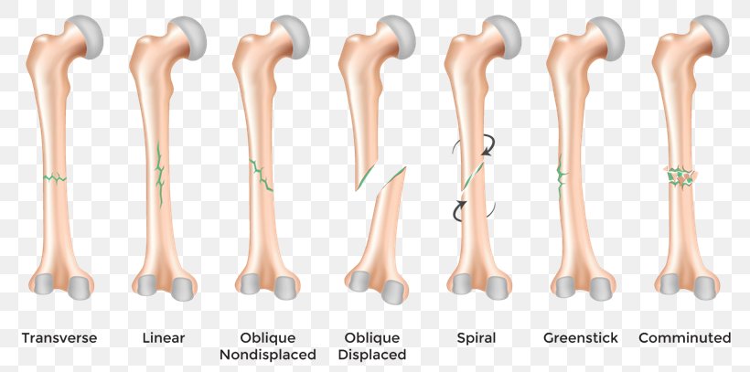
Bone fracture diagram femoral fracture femur, png, 810x407px ...
The anatomy of femoral vascular access — taming the sru
Femoral artery - location, anatomy, branches, function and faqs
Locations for pin placement in the femur
0 Response to "40 where on the diagram is the femoral area?"
Post a Comment