36 nerves of the foot and ankle diagram
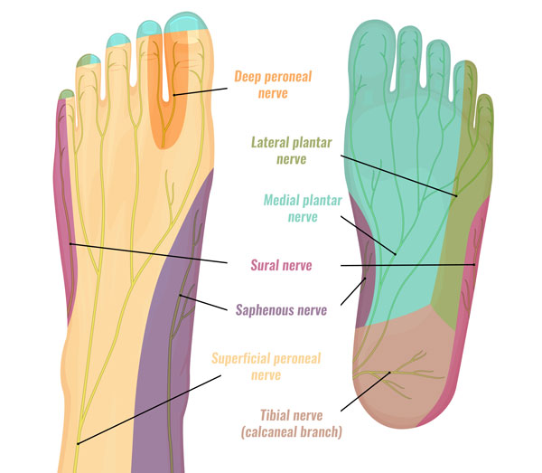
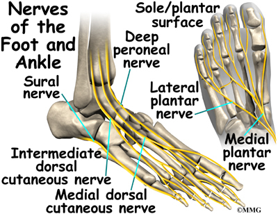
The nerves of the foot assistance move the body and keep balance both while it’s moving and at rest. All these nerves extend as branches of nerves in the leg that travel through the ankle and into the foot. The sural nerve branches from the tibial and common fibular nerves and is responsible for feeling on the outside of the foot and the little toe. The median and lateral plantar nerves are the 2 biggest nerves in the bottom of the foot. Jul 03, 2018 · All of these nerves extend as branches of nerves in the leg that pass through the ankle and into the foot. The sural nerve branches from the tibial and common fibular nerves and is responsible for feeling on the outside of the foot and the small toe. The medial and lateral plantar nerves are the two largest nerves in the bottom of the foot.
The innervation of the lower leg, ankle and foot begins upstream at the lombo-sacral region of the spine with a bundle of nerves that together form the lumbo-sacral plexus. The nerves exiting the 4th and 5th lumbar vertebrae form the lumbo-sacral trunk merge with the sacral plexus, which consists of the sacral nerves from the 1st to the 4th ...

Nerves of the foot and ankle diagram
Damage to the foot and ankle tendons are a common cause of foot pain, typically caused by overuse, overstretching or an injury. Tendons are thick bands of tissue that connect muscles to bone. When a muscle contracts, the tendon pulls on the bone causing the joint to move. There are a number of tendons located in the foot and ankle all ... various nerves about the ankle and foot. It is also important to recognize common anatomic vari-ants. In this article, we review the normal anat-TEACHING POINTS During ankle dorsiflexion, excessive traction may occur along the exit point of the superficial peroneal nerve, injuring it and causing secondary neuroma formation at this level. The nerve Foot Pain Diagram. Written By: Chloe Wilson BSc(Hons) Physiotherapy Reviewed By: FPE Medical Review Board A foot pain diagram is a great tool to help you work out what is causing your ankle and foot pain. There are a whole range of structures e.g. bones, muscles, tendons and nerves which will each give slightly different foot pain symptoms.
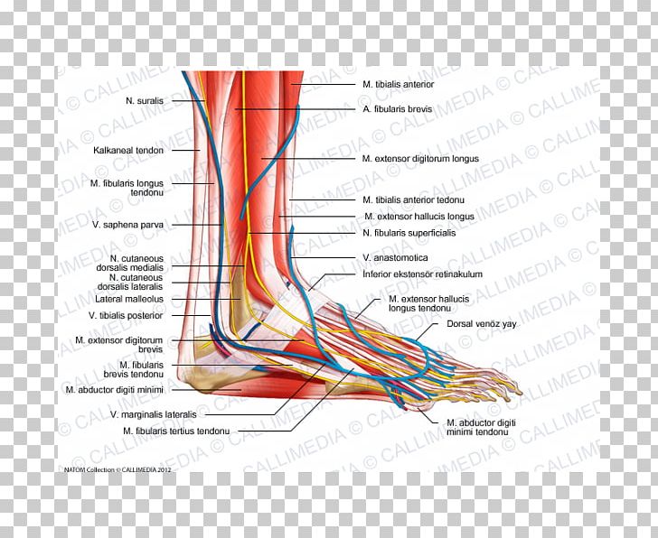
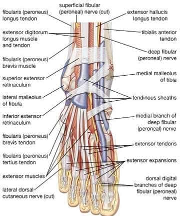
Nerves of the foot and ankle diagram. Nerves In Foot Diagram. nerves of the leg and foot along its route through the legs the sciatic nerve splits into the tibial and mon fibular peroneal nerves which in turn split into many smaller nerves in the legs and feet the nerves of the foot help move the body and keep balance both while it’s moving and at rest a plete guide to the nerves in your feet foot vitals tingling feet the “pins and needles” sensation also known as paresthesia is a very mon symptom find out causes ... Problems with nerves in the feet are very common. Many times, an injured nerve will cause intense pain and heat felt within the foot. Nerves act as a network, communicating important information from the foot to the brain. Learn more about the various conditions and problems that can affect the nerves in the foot. The anatomy of the nerves of the foot and ankle is complex, and familiarity with the normal anatomy and course of these nerves as well as common anatomic variants is essential for correct identification at imaging. Ultrasonography (US) and magnetic resonance (MR) imaging allow visualization of these nerves and may facilitate diagnosis of various compression syndromes, such as "jogger's ... The deep fibular nerve is parallel and lateral to the tendon of the extensor hallucis longus muscle and goes inside the dorsal aspect of the foot on the lateral aspect of the dorsalis pedis artery. The nerve produces a lateral branch just distal towards the ankle joint, which stimulates the extensor digitorum brevis from its deep surface. The ...
The anatomy of the foot. The foot contains a lot of moving parts - 26 bones, 33 joints and over 100 ligaments. The foot is divided into three sections - the forefoot, the midfoot and the hindfoot. The forefoot. This consists of five long metatarsal bones and five shorter bones that form the toes (phalanges). The midfoot Nerves of the Foot. There are five main nerves that run past the ankle into the foot (Figure 17). All five of these are derived from two nerves that originate from the lumbar spine. The sciatic nerve branches into four of the five primary nerves of the foot. Two segments of the sciatic nerve branch before the knee joint: the tibial nerve and peroneal nerve. by M De Maeseneer · 2015 · Cited by 70 — The anatomy of the nerves of the foot and ankle is complex, and familiarity with the normal anatomy and course of these nerves as well as common anatomic ... Dr. Ebraheim's educational animated video describes the nerves of the lower leg in a very easy and simple animation.Lateral cutaneous nerve of the calfSural ...
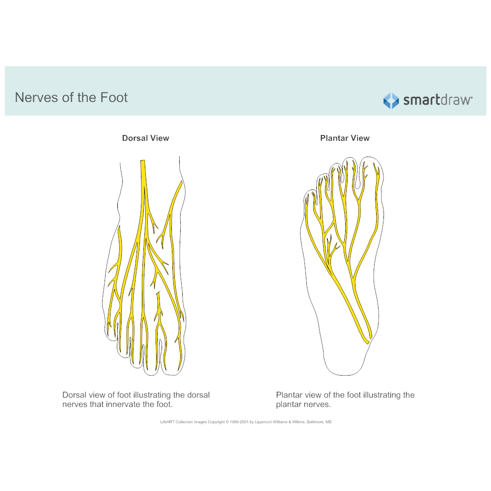
Nerves in the feet send messages, such as indications of heat and pain and other information, to the brain. The dorsal digital nerves of the foot branch throughout the body of the foot and down ... There are six nerves associated with the motor and sensory functions of the foot and ankle. They are: Nerve Innervations: Superficial peroneal nerve (L4-S1) Lateral plantar nerve (S2-S3) Medial plantar nerve (L5-S3) This nerve enters the foot along the outside of the ankle and runs to the fifth toe, supplying only the lateral side of the toe. On the opposite side of the foot is a nerve that is not counted among the dorsal digital nerves, the saphenous nerve. This nerve penetrates the medial side of the foot, ending just beneath the big toe, and it is the ... Match the corresponding numbers on the foot diagram below for a list of conditions that may be causing your foot and ankle pain. This is meant for educational purposes only. If you're having a problem with your foot or ankle, visit a podiatrist - a foot and ankle specialist! Top (Dorsal) View of Foot & Ankle Number 1 and 2:
Nerves Of The Leg & Foot - Everything You Need To Know ...
3%. (70/2732) 3. It is the terminal branch of the superficial peroneal nerve; injury leads to reduced sensation over medial aspect of great toe. 83%. (2260/2732) 4. It is the terminal branch of the deep peroneal nerve; injury leads to first interphylangeal joint flexion weakness. 3%.

Warm bath before treatment
Extensor digitorum brevis, Origin: Superolateral surface of calcaneus bone. Insertion: Middle phalanges of toes 2-4. Innervation: Deep fibular/peroneal nerve ( ...Ankle anatomy: Movements: dorsiflexion, plant...Bones of the foot: Tarsals: proximal (talus, calc...Muscles of the foot: Movements: foot inversion, ...Joints of the foot: Intertarsal: subtalar (talocalc...

Foot Tendon Anatomy Diagram | Ankle tendonitis, Ankle ...
by A Tang · 2021 · Cited by 1 — The foot receives its nerve supply from the superficial peroneal (fibular) nerve, deep fibular nerve, tibial nerve (and its branches), ...

One and one — is three.
Neuritis and Neuromas of the Foot and Ankle. Edited by Eric Malicky MD . Clinical Presentation. Injury or irritation to nerves in the foot and ankle often creates pain and/or numbness. They occur as a result of an injury to a specific nerve. Nerve damage can result from the original injury, casting/wrap, or surgery.
Foot Nerve Human Anatomy Muscle PNG, Clipart, Anatomy ...
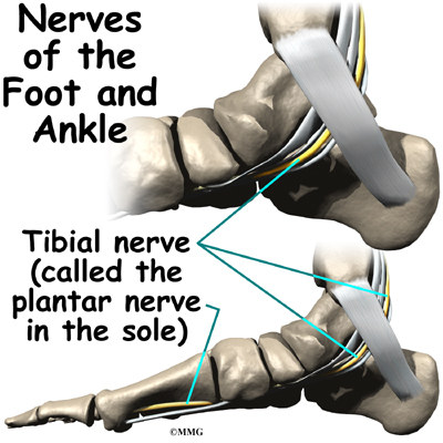
6 Jul 2020 — The main nerve to the foot, the posterior tibial nerve, enters the sole of the foot by running behind the inside bump on the ankle (medial ...

http://slideplayer.com/slide/9411428/28/images/27/Nerves ...
The nerves of the ankle are derived from the deep and superficial peroneal nerves, the tibial nerves, and the sural and saphenous nerves. The Foot Bones of the foot as seen from the medial (arch) side. The foot is a firm platform that support the weight of the body.

EM Didactic: Ankle Block (Landmark Technique) - Procedures
Foot Pain Diagram. Written By: Chloe Wilson BSc(Hons) Physiotherapy Reviewed By: FPE Medical Review Board A foot pain diagram is a great tool to help you work out what is causing your ankle and foot pain. There are a whole range of structures e.g. bones, muscles, tendons and nerves which will each give slightly different foot pain symptoms.
Medial Calcaneal Nerve Entrapment - Symptoms & Treatment
various nerves about the ankle and foot. It is also important to recognize common anatomic vari-ants. In this article, we review the normal anat-TEACHING POINTS During ankle dorsiflexion, excessive traction may occur along the exit point of the superficial peroneal nerve, injuring it and causing secondary neuroma formation at this level. The nerve

Figure 1 from Peripheral nerve crushing to relieve chronic ...
Damage to the foot and ankle tendons are a common cause of foot pain, typically caused by overuse, overstretching or an injury. Tendons are thick bands of tissue that connect muscles to bone. When a muscle contracts, the tendon pulls on the bone causing the joint to move. There are a number of tendons located in the foot and ankle all ...

Diagram Showing Parts Of The Foot
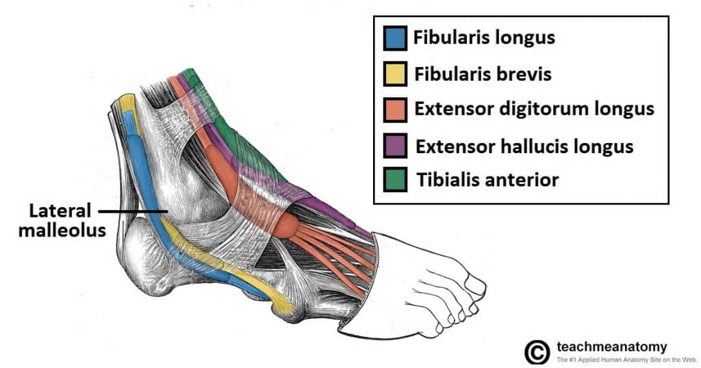
Muscles in the Lateral Compartment of the Leg - TeachMeAnatomy

Frog Sculpture, Dunston, Gateshead, Tyne & Wear, England.

Sandy feet at beach

nerves in the feet diagram - Google Search | Injury Help ...
Nerves of the Foot

An infant’s feet

Tarsal Tunnel Syndrome - Foot & Ankle - Orthobullets

Image from page 85 of "The American journal of anatomy" (1905)

Image from page 79 of "The treatment of fractures" (1901)
Ankle Anatomy | eOrthopod.com

Image from page 88 of "An illustrated encyclopædic medical dictionary. Being a dictionary of the technical terms used by writers on medicine and the collateral sciences, in the Latin, English, French and German languages" (1890)

Nerves of foot. (adapted from Encyclopedia Britanica 2007 ...

Nerves of the Foot | Fodterapeut | Pinterest

Barefoot at the beach

Frog, Deep Sea World, North Queensferry, Fife, Scotland.
Lower limb arteries and nerves: Anatomy, branches | Kenhub

Dry Docked – Three Steps to Avoiding Ankle Injuries ...
A Patient’s Guide to Ankle Anatomy | Houston Methodist

Pin on Feet Care
anatomical landmarks on the dorsum of the left foot ...

Areas of the foot and their associated nerves: (1 ...
Nerves in Foot and Ankle | IYTmed.com

Live simple

Several things about Anatomy of the Hip Muscles | Foot ...

Foot pain nerve, how to get rid of corns on toes in a week

After the accident
0 Response to "36 nerves of the foot and ankle diagram"
Post a Comment