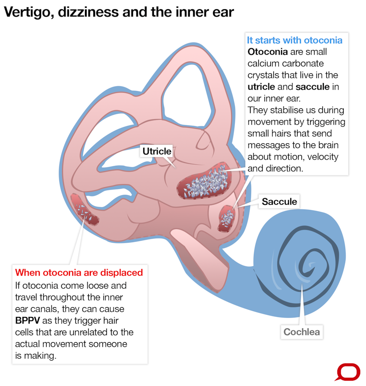
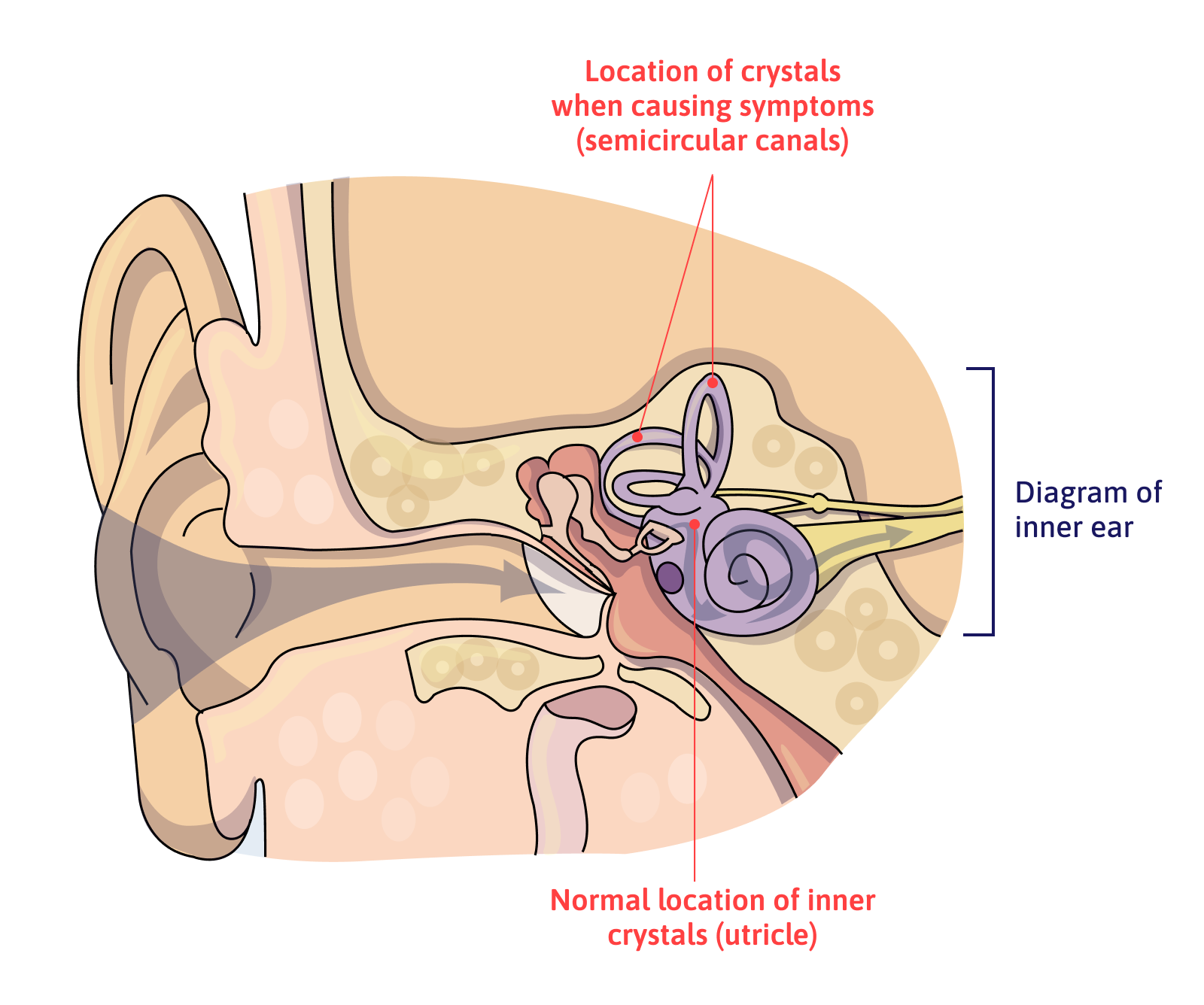
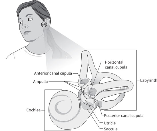
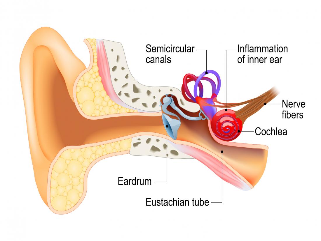
36 inner ear crystals diagram
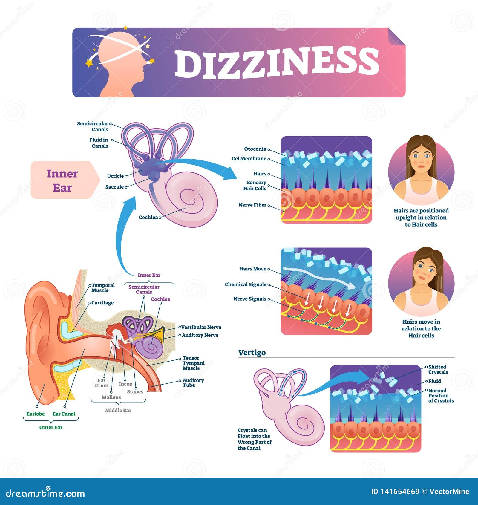
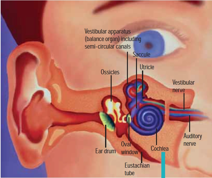
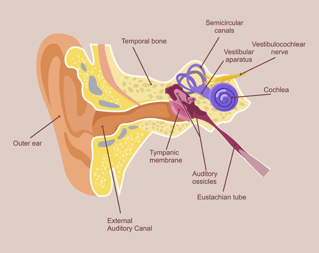
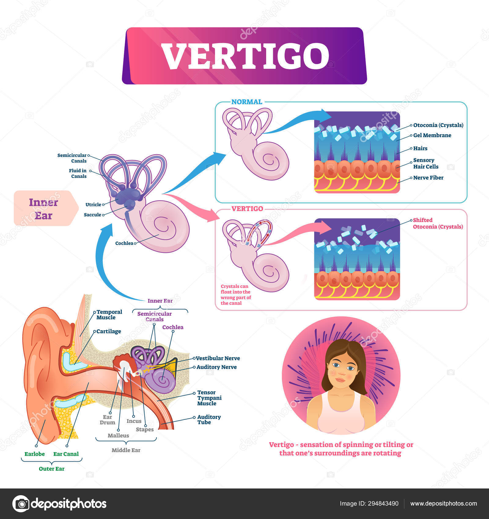
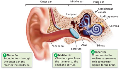
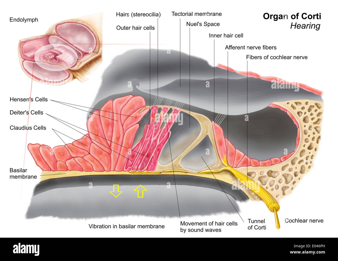
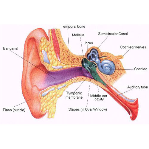
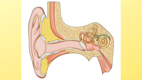
Diagram of outer, middle, and inner ear. ... (The rest of the inner ear, that is, the cochlea, is concerned with hearing.) ... The treatment of vertigo may include medication, special exercises to reposition loose crystals in the inner ear, or exercises designed to help the patient re-establish a sense of equilibrium. Controlling risk factors ... Browse 1,560 inner ear stock photos and images available, or search for inner ear illustration or inner ear diagram to find more great stock photos and pictures. Illustration of hearing, journey of the sound wave in the ear. The sound wave is captured by the auricle, penetrates in the auditory canal, vibrates...
Aug 06, 2016 · BPPV is a result of tiny crystals in your inner ear being out of place. The crystals make you sensitive to gravity and help you to keep your balance. Normally, a jelly-like membrane in your ear keeps the crystals where they belong. If the ear is damaged — often by a blow to the head — the crystals can shift to another part of the ear.

Inner ear crystals diagram
Jan 29, 2019 · When they are dislodged, the crystals float around in the fluid area of the balance branch of the inner ear, and you will start to feel off balance. The loose crystals will start to make people feel like they are spinning and the room is spinning around them. If you are 60 or older, you are more prone to having your ear crystals dislodge. One Ear from nest 1 to nest 2 (short left, long right). The Camoudile falls in the pit. Rabbit Ears from nest 2 to nest 5 (long right, short middle). Whitey from nest 4 to nest 5 (long left, long middle). Flat head from nest 3 to nest 4 (short middle, short left). One Ear from nest 2 to nest 3 (short left, long right). The vestibular system is located within the inner ear. Laterally, it is bordered by the middle ear and medially, ... Diagram of the inner ear. Source: Blausen.com staff (2014). ... contains calcium carbonate crystals called otoconia. These crystals play an important role in bending hair cells (and therefore, detecting linear acceleration ...
Inner ear crystals diagram. Dec 21, 2021 · Vestibular system anatomy. The vestibular system is a somatosensory portion of the nervous system that provides us with the awareness of the spatial position of our head and body (proprioception) and self-motion (kinesthesia).). It is composed of central and peripheral portions. The peripheral portion of the vestibular system consists of the vestibular labyrinth, … Dec 14, 2021 · The blades of grass represent cilia, hair-like processes that are attached to tiny nerves in your inner ear. When the crystals move, it stimulates the nerves to fire, which tells the brain your ... Diagram of the inner ear with the semicircular canals highlighted. Causes. Causes of BPPV can be: spontaneous – no cause, with the risk increasing with age; trauma; for example, after a knock to the head or concussion; recent inner ear infection. Diagnosis BPPV is caused by a problem in your inner ear. Your semicircular canals are found inside your ear. They detect motion and send this information to your brain. The utricle is a nearby part of the ear. It contains calcium crystals (canaliths) that help it detect movement.
Eyes are organs of the visual system.They provide living organisms with vision, the ability to receive and process visual detail, as well as enabling several photo response functions that are independent of vision.Eyes detect light and convert it into electro-chemical impulses in neurons.In higher organisms, the eye is a complex optical system which collects light from the surrounding ... Diagram of outer, middle, and inner ear. The outer ear is labeled in the figure and includes the ear canal. ... The calcium crystals are the structures that ultimately stimulate the position hairs and provoke nerve impulses created by the position changes and transmit that information to the brain stem and cerebellum. Feb 03, 2022 · For these earrings you need superduos, four 4mm crystals, and two 6mm crystals (round or bicone). You will also need 11/0 seed beads plus two 6/0 or 8/0 seed beads for the jump rings to go through to connect the ear hooks or clips. The 4mm crystals are for the top and middle of the trees, and can be the same or different colors. Jul 29, 2020 · Found in a small cavity inside of the temporal bone, they serve to transmit and amplify sound from the eardrum to the inner ear. Vertebrae. Twenty-six vertebrae form the vertebral column of the human body. They are named by region: Cervical (neck) - 7 vertebrae; Thoracic (chest) - 12 vertebrae; Lumbar (lower back) - 5 vertebrae; Sacrum - 1 vertebra
Human Ear: Structure and Functions (With Diagram) ... of the inner ear. Malleus is the largest ossicle, however, stapes is smallest ossicle. Stapes is also the smallest bone in the body. ... the otolith membrane in which there are also found very small crystals of calcium carbonate, the otolith. The cristae and maculae are the receptors of balance. The vestibular system is located within the inner ear. Laterally, it is bordered by the middle ear and medially, ... Diagram of the inner ear. Source: Blausen.com staff (2014). ... contains calcium carbonate crystals called otoconia. These crystals play an important role in bending hair cells (and therefore, detecting linear acceleration ... One Ear from nest 1 to nest 2 (short left, long right). The Camoudile falls in the pit. Rabbit Ears from nest 2 to nest 5 (long right, short middle). Whitey from nest 4 to nest 5 (long left, long middle). Flat head from nest 3 to nest 4 (short middle, short left). One Ear from nest 2 to nest 3 (short left, long right). Jan 29, 2019 · When they are dislodged, the crystals float around in the fluid area of the balance branch of the inner ear, and you will start to feel off balance. The loose crystals will start to make people feel like they are spinning and the room is spinning around them. If you are 60 or older, you are more prone to having your ear crystals dislodge.











0 Response to "36 inner ear crystals diagram"
Post a Comment