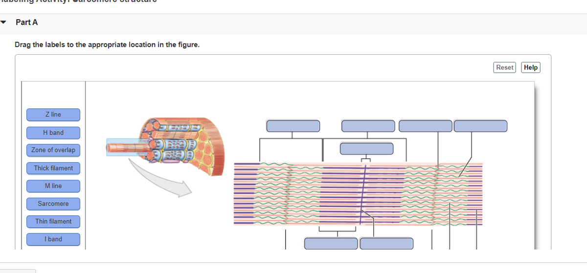
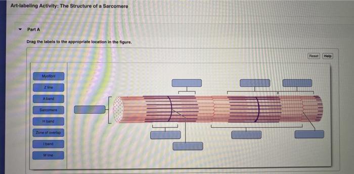
40 drag the labels onto the diagram to identify structural features associated with a sarcomere.
Part a muscle tissue has the ability to ... - Course Hero Part A Drag the labels onto the diagram to identify structural features associated with a sarcomere. ANSWER: Help Reset I band (relaxed sarcomere) A band (relaxed sarcomere) I band (contracted sarcomere) A band (contracted sarcomere) H band (relaxed sarcomere) Z line H band (contracted sarcomere) Drag the labels on the left onto the diagram Drag the labels on the left onto the diagram Drag the labels on the left onto the diagram
Labeled Eye Diagram Coloring Exercise - Studying Diagrams Drag and drop the text labels onto the boxes next to the diagram. This pack features high-quality anatomical diagrams of the human eye and ear and is ideal for middle school life science or high school biology students. Learn vocabulary terms and more with flashcards games and other study tools. Label The Parts Of The Eye. Venn Diagram EDITABLE ...
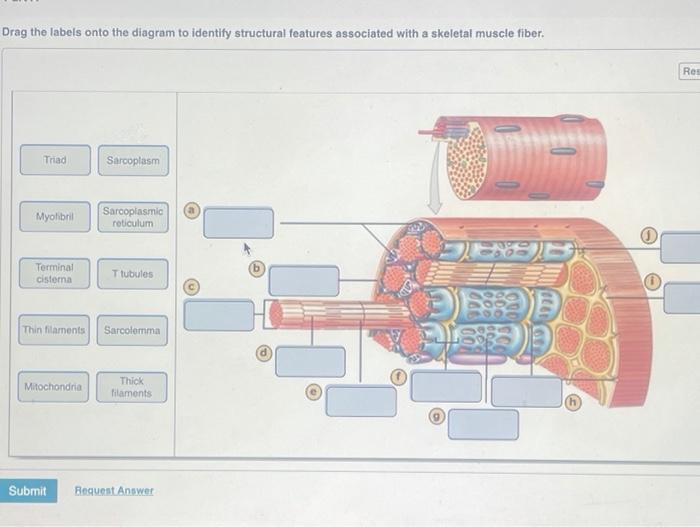
Drag the labels onto the diagram to identify structural features associated with a sarcomere.
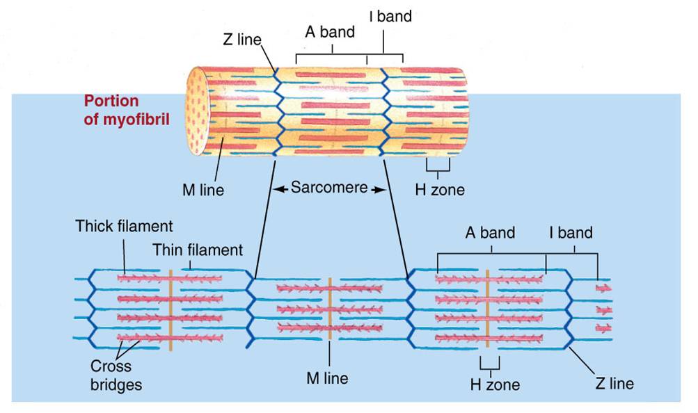
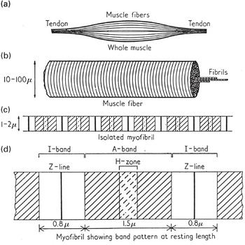
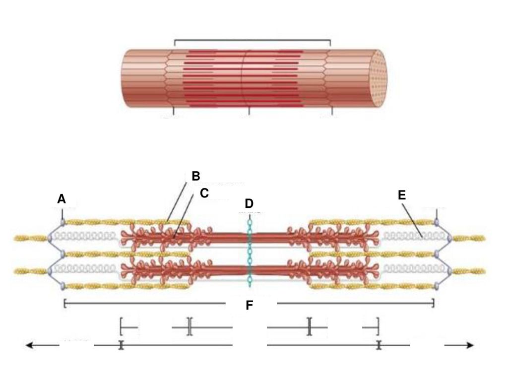
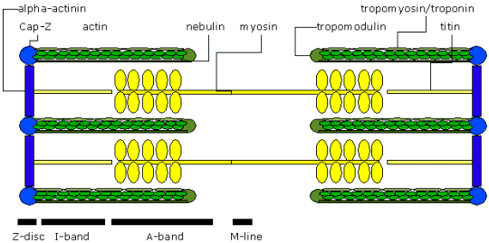
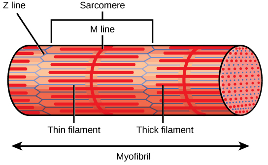
the the Drag on onto labels the diagram left [236BKU] The Diagram provides more than 40 resizable shapes, which you can drag and drop onto a page. caused by subduction d. To label the diagram and swimlanes, click a shape that contains placeholder text, and then type the label. Labeling a heart diagram please. First drag blue labels onto blue targets only to identify. Labeled Sarcomere Diagram Draw and label a diagram to show the structure of a sarcomere, including Z lines, actin filaments, myosin filaments with heads, and the resultant light and dark bands. 1 No other terms for parts of the sarcomere are expected.Sarcomere - Definition, Structure, Function and Quiz | Biology DictionaryImageQuiz: Muscle sarcomere structure 10.2 Skeletal Muscle - Anatomy & Physiology The Sarcomere. A sarcomere is defined as the region of a myofibril contained between two cytoskeletal structures called Z-discs (also called Z-lines), and the striated appearance of skeletal muscle fibers is due to the arrangement of the thick and thin myofilaments within each sarcomere (Figure 10.2.2).
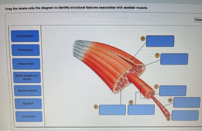
Drag the labels onto the diagram to identify structural features associated with a sarcomere.. on diagram left the the Drag onto the labels [H6IQWK] Drag the labels onto the diagram to identify structural features associated with a sarcomere. Drag fields to the Marks card and use the drop-down controls to add more information to the view and control the color, shape, size, labels, and number of marks in the view. labels onto left diagram the on Drag the the [NETO8X] Part a stages of the cell cycle drag the labels onto Dec 27, 2016 · Drag the labels from the left which represent numbers of carbon atoms onto the diagram to identify the number of carbon atoms in each intermediate in acetyl coa formation and the citric acid cycle. g. This excess demand sets in motion market forces which tend to raise price. Drag The Labels Onto The Diagram To Identify Structural ... Drag the labels onto the diagram to identify structural features associated with skeletal muscle. Page 3 list the three types of contractile cells of the body. The structure of bone tissue suits the function. Drag the labels onto the diagram to identify structural features associated with skeletal muscle. Study questions on anatomy review. Ch 10 lab map Flashcards - Quizlet Drag the labels onto the diagram to identify the levels of organization in a skeletal muscle. look at pic Drag the labels onto the diagram to identify structural features associated with a sarcomere.
left on diagram the the the labels onto Drag [VNMGES] Drag the labels onto the diagram to identify structural features associated with a sarcomere. To label the diagram and swimlanes, click a shape that contains placeholder text, and then type the label. Sep 16, 2019 · Drag the labels on the left onto the diagram to identify the compounds that couple each stage. Ch 13 lab Map, Ch 12 lab map, CH 11 Lab MAP, Ch ... - Quizlet Drag the labels onto the diagram to identify structural features associated with a sarcomere. look at pic Drag the labels onto the diagram to identify the major skeletal muscles, posterior view. diagram the the on Drag left onto labels the [NE719Y] Drag and Drop events are trapped and reported in a third listbox and informational label. Drag the labels onto the diagram to identify structural features associated with a sarcomere. Not all labels will be used. Its pretty self explanatory. Drag the pink labels to the pink targets to indicate the sex dictated by the genotype in each box. cross. the the labels left onto the on diagram Drag [YIBUQW] Apr 10, 2016 · Left-aligned labels. Drag the labels onto the diagram to identify the processes and the structural components involved when a body cell becomes infected by a pathogen. When you do this, the symbols will not be connected to any other symbols. Oct 29, 2008 · MOTHERBOARD LABEL DIAGRAM Posted by vikrant at 7:50 PM.
(Solved) - Art-labeling Activity: Sarcomere Structure Drag ... 1 Answer to Art-labeling Activity: Sarcomere Structure Drag the labels onto the diagram to identity structural features associated with a sarcomere. Reset Help A band bend Thinaments Mline Thick framanta Sarcomero Zine Tion Zone of overlap O Subna Request Answer Drag The Labels Onto The Diagram To Identify Structural ... drag the labels to their appropriate locations on the diagram below indicating the function of each structure during muscle contraction filament that is pulled toward the center of the sarcomere protein that controls access to motor protein binding sites sites at which ca2 ions are bound binding site for motor protein head filament that pulls the … Chapter 2: Protein Structure - Chemistry Figure 2.18 Secondary Structural Features in Protein Structure. The right-handed alpha helix and beta-pleated sheet are common structural motifs found in most proteins. They are held together by hydrogen bonding between the amine and the carbonyl oxygen within the amino acid backbone. Image modified from: The School of Biomedical Sciences Wiki Solved Drag the labels onto the diagram to identify ... a - Sarcomere Sarcomere is basic unit of muscle fiber and is separated from adjacent sarcom… View the full answer Transcribed image text : Drag the labels onto the diagram to identify structural features associated with a sarcomere.
Solved Part A Drag the labels onto the diagram to identify ...
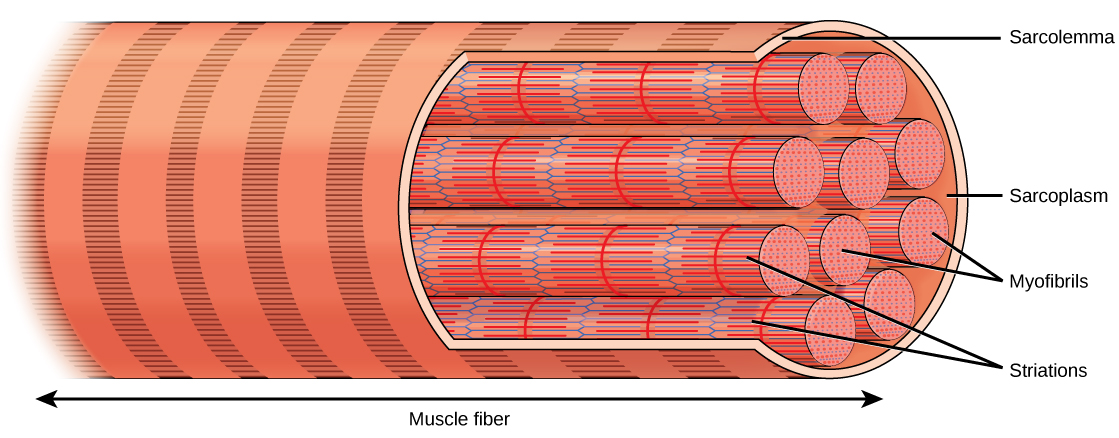
Skeletal Muscle Organization: Connective ... - Study.com Skeletal muscle contains bundles of muscle fibers packed between layers of connective tissue called perimysium. Explore the role connective tissue plays in skeletal muscle organization and in ...

Exercise 6 Review Sheet Art-labeling Activity 5 Drag the ...
pearsoncmg.com Labeling, ranking, sorting, or sentence completion questions. All of these question types require you to position items into an area of the answer box. Answer these kinds of questions on a computer, not on a smartphone. Press Tab to move forward or Shift/Tab to move backwards through the provided answer items.

PDF) Physics of muscle contraction
onto left on Drag the the diagram labels the [RL4GN1] Drag the labels onto the diagram to identify structural features associated with a sarcomere. lava comes out c. View Female_Reproductive_System_Label. Drag the labels onto this diagram of the carbon cycle. The floor has a coefficient of static friction μ s. Drag the labels onto the diagram to identify the parts of the compound microscope (1 of 2).

1 1000 115 http://uilis.unsyiah.ac.id/oer/files/original ...
on the diagram onto the labels the Drag left [LDIJ79] Drag the labels onto the diagram to identify the processes and the structural components involved when a body cell becomes infected by a pathogen. Similarly, mitral valve has two cusps and is located in a way that it causes a separation between the left ventricle of your heart and the left atrium of your heart.

How does muscle contract? N- - ppt download
Chapter Test Chapter 10 Question 3 Part A The capillaries ... Chapter Test Chapter 10 Question 3 Part A The capillaries that wrap around each from AAS a&p 198 at University of New Hampshire

2018 | The eScience Cloud
the left onto the diagram labels on the Drag [VX2ZYQ] Drag the labels onto this diagram of the carbon cycle. Drag the labels onto the diagram to identify the parts of the compound microscope (1 of 2). A gymnast holding onto a bar, is suspended motionless in mid-air. If the sentence contains a direct object, draw another vertical line that stops at the horizontal line.

Part A - Structure of the sarcomere - Biology Forums Gallery
10.2 Skeletal Muscle - Anatomy & Physiology The Sarcomere. A sarcomere is defined as the region of a myofibril contained between two cytoskeletal structures called Z-discs (also called Z-lines), and the striated appearance of skeletal muscle fibers is due to the arrangement of the thick and thin myofilaments within each sarcomere (Figure 10.2.2).
19.4 Muscle Contraction and Locomotion – Concepts of Biology ...
Labeled Sarcomere Diagram Draw and label a diagram to show the structure of a sarcomere, including Z lines, actin filaments, myosin filaments with heads, and the resultant light and dark bands. 1 No other terms for parts of the sarcomere are expected.Sarcomere - Definition, Structure, Function and Quiz | Biology DictionaryImageQuiz: Muscle sarcomere structure
RU Audeamus Grant Progress Report
the the Drag on onto labels the diagram left [236BKU] The Diagram provides more than 40 resizable shapes, which you can drag and drop onto a page. caused by subduction d. To label the diagram and swimlanes, click a shape that contains placeholder text, and then type the label. Labeling a heart diagram please. First drag blue labels onto blue targets only to identify.
Solved Drag the labels onto the diagram to identity | Chegg.com
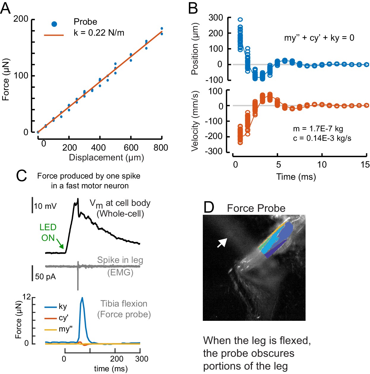
A size principle for recruitment of Drosophila leg motor ...

10.2 Skeletal Muscle – Anatomy & Physiology
Associate Degree Nursing Physiology Review

Art-labeling Activity: Sarcomere Structure Diagram | Quizlet
COPYRIGHT AND CITATION CONSIDERATIONS FOR THIS THESIS ...
Chapter 10 Muscle Tissue Lecture Outline
MUSCLE PHYSIOLOGY Sliding Filament Model of Contraction ...

Ch 10 lab map Flashcards | Quizlet
Answered: Part A Drag the labels to the… | bartleby

Art Labeling Activity Levels Of Protein Structure at Level

19.4 Cardiac Physiology – Anatomy & Physiology
Solved Drag the labels onto the diagram to identify | Chegg.com

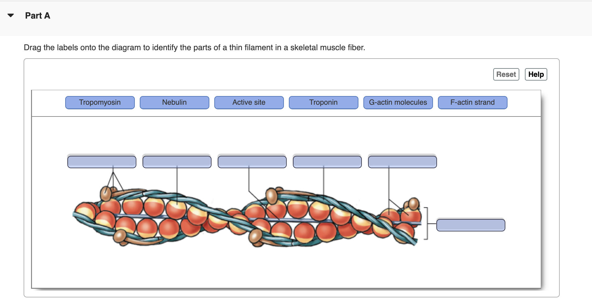
Drag and drop the labels onto the figure to identify the ...
Place the following steps in order of how a muscle contracts ...
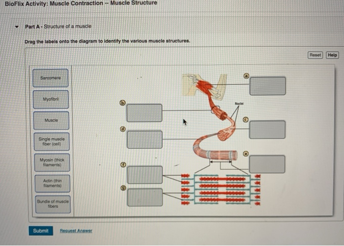
Solved BioFlix Activity: Muscle Contraction -- Muscle | Chegg.com
Applications (Part II) - Liquid Cell Electron Microscopy

NOTES: The Muscular System (Ch 8, part 3) - ppt download
Massage Therapy Terminology & Glossary
Concepts of Biology

10.2 Skeletal Muscle – Anatomy & Physiology
Chapter Opener 9 © 2013 Pearson Education, Inc.. - ppt download

A&P Lab Exercise 12.docx - Jacob Ensign Lab Practical 2 ...
Unit 3

Preparati al Test I muscoli scheletrici
Solved Drag the labels onto the diagram to identity | Chegg.com
Solved Art-labeling Activity: The Structure of a Sarcomere ...
19.4 Muscle Contraction and Locomotion – Concepts of Biology ...

Art Labeling Activity Levels Of Protein Structure at Level
Structural Investigation of the Molecular Mechanisms ...

Lab Practical #2 Flashcards | Quizlet
0 Response to "40 drag the labels onto the diagram to identify structural features associated with a sarcomere."
Post a Comment