36 diagram of synovial joint
The Six Types of Synovial Joints: Examples & Definition ... This type of joint can be found between your neck vertebrae. For instance, when you turn your head side-to-side, it's due to the rotary motion permissible in pivot joints. Next, let's focus on hinge joints, shown as letter B on the diagram. Hinge joints are the synovial joint type referred to in our introductory section. Dog Leg Anatomy with Labeled Diagram - Bones, Joints ... 07-09-2021 · Dog leg anatomy. First, you might have a basic idea of the different bones of the forelimb and hindlimb of a dog. Now I will provide you the few information on the other bones of dog leg anatomy with their unique features. The front leg of a dog consists of the clavicle, scapula (arm), radius and ulna (forearm), carpals, metacarpals, and phalanges (forepaw).
Synovial Joints - Anatomy and Physiology synovial joint at which the convex surface of one bone articulates with the concave surface of a second bone; includes the elbow, knee, ankle, and interphalangeal joints; functionally classified as a uniaxial joint. intracapsular ligament. ligament that is located within the articular capsule of a synovial joint.
Diagram of synovial joint
Synovial Joints Anatomy Diagram - Quizlet The only 2 synovial joints that aren't diarthrotic. carpals and tarsals. Functional classification of carpals and tarsals. amphiarthrosis. membrane continuous from bone to bone outside the articular capsule. periosteum. fibrous capsule lined by synovial membrane. articular capsule. Structures of a Synovial Joint - Capsule - Ligaments ... A synovial joint is characterised by the presence of a fluid-filled joint cavity contained within a fibrous capsule. It is the most common type of joint found in the human body, and contains several structures which are not seen in fibrous or cartilaginous joints.. In this article we shall look at the anatomy of a synovial joint - the joint capsule, neurovascular structures and clinical ... Synovial joints - Skeletal system - AQA - GCSE Physical ... Synovial joints. A joint is a place where two or more bones meet and is also called an articulation. The role of joints and connective tissue . Connective tissues consist of ligaments, cartilage ...
Diagram of synovial joint. Sacroiliac joint: Anatomy, function | Kenhub The sacroiliac joint is a synovial joint formed between the ilium and the sacrum. The left and right sacroiliac joints, together with the pubic symphysis and the sacrococcygeal joint, compose the articulations of the pelvic girdle. The sacroiliac joints connect the hip bones posterolaterally, while the pubic symphysis connects them anteriorly. Synovial Joints | Anatomy and Physiology I The six types of synovial joints allow the body to move in a variety of ways. (a) Pivot joints allow for rotation around an axis, such as between the first and second cervical vertebrae, which allows for side-to-side rotation of the head. (b) The hinge joint of the elbow works like a door hinge. (c) The articulation between the trapezium carpal ... Types of Joints in Animals with Example and Diagrams ... Synovial joint diagram Here in the diagram, you will find all the structures of a synovial joints in animals. If you need more diagram like this synovial joint then, you may follow anatomy learner blog or social media. Again, you might read other different article related to veterinary osteology or syndesmology with the anatomy learner. Synovial Joints.docx - -Synovial Joints-USLOs ... -----Synovial Joints-----USLOs: Differentiate the structure of 6 synovial joints and tell their different functions/movements. Give an example of each. Synovial Joints: joint at which the articulating surfaces of the bones are located within a joint cavity formed by an articular capsule Most common type of joint in the body Only one to have a joint cavity (not in fibrous or cartilaginous) The ...
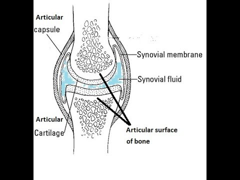
Saddle joint - Wikipedia A saddle joint (sellar joint, articulation by reciprocal reception [citation needed]) is a type of synovial joint in which the opposing surfaces are reciprocally concave and convex. It is found in the thumb , the thorax , the middle ear , and the heel . Synovial Joint: structure and label Diagram | Quizlet Encloses the joint cavity Consists of 2 parts: 1. Fibrous Capsule (outer)- provides joint stability 2. Synovial Membrane (inner)- secretes (produces) synovial fluid for lubrication Classification of Joints | Boundless Anatomy and Physiology Synovial Joint: This diagram of a synovial joint delineates the articular cartilage, articular capsule, bone, synovial membrane, and joint cavity containing synovial fluid. Synovial Joints This is the most common and movable joint type in the body. The Six Types of Synovial Joints: Examples & Definition ... Next, let's focus on hinge joints, shown as letter B on the diagram. Hinge joints are the synovial joint type referred to in our introductory section. These joints can be found between your upper ...
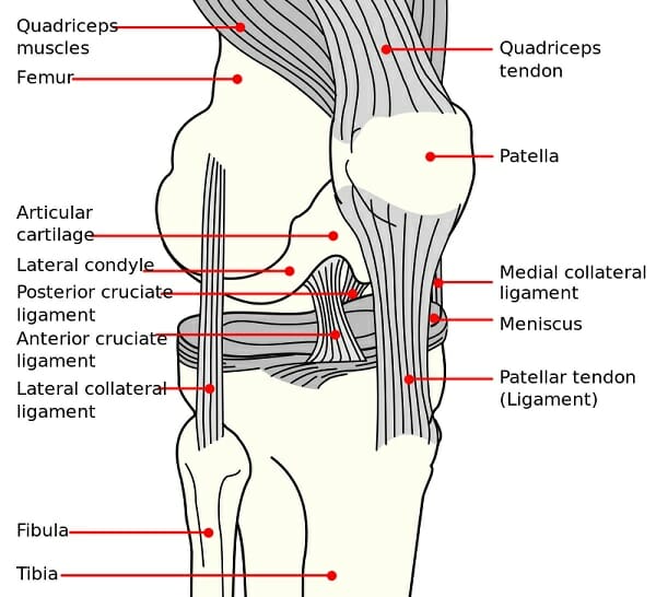
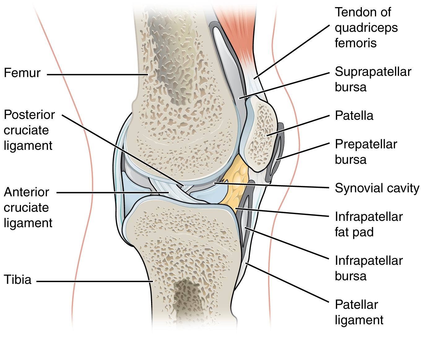
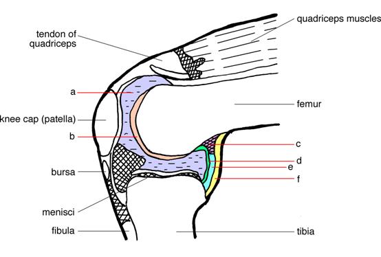
Ankle - Wikipedia The talocrural joint is a synovial hinge joint that connects the distal ends of the tibia and fibula in the lower limb with the proximal end of the talus. The articulation between the tibia and the talus bears more weight than that between the smaller fibula and the talus. Synovial Joints | Boundless Anatomy and Physiology A synovial membrane (or synovium) is the soft tissue found between the articular capsule (joint capsule) and the joint cavity of synovial joints. Synovial fluid is the clear, viscid, lubricating fluid secreted by synovial membranes. The morphology of synovial membranes may vary, but it often consists of two layers. Labelled Diagram Of Synovial Joint - Wiring Diagrams The basic structure of a synovial joint is shown in the diagram on the right. The main parts of synovial joints are labelled on the synovial joint diagram and described in the table below. Some synovial joints are more complicated than others. An example of a simple synovial joint, e.g. a metacarpophalangeal (finger) joint, is shown above-right ... The Knee Joint - Articulations - Movements - Injuries ... Feb 07, 2022 · The knee joint is a hinge type synovial joint, which mainly allows for flexion and extension (and a small degree of medial and lateral rotation). It is formed by articulations between the patella, femur and tibia. In this article, we shall examine the anatomy of the knee joint – its articulating surfaces, ligaments and neurovascular supply.
Elbow synovial joint - Labelled diagram
Bones Joints and Cartilage Notes: Diagrams & Illustrations ... This Osmosis High-Yield Note provides an overview of Bones Joints and Cartilage essentials. All Osmosis Notes are clearly laid-out and contain striking images, tables, and diagrams to help visual learners understand complex topics quickly and efficiently. Find more information about Bones Joints and Cartilage by visiting the associated Learn Page.
Synovial Joint (Diarthrosis): Definition, Types, Structure ...
[SOLVED] Draw a labelled diagram of a synovial joint. Give ... Draw a labelled diagram of a synovial joint. Give one example each for a hinge joint, a pivot joint, axial skeleton and appendicular skeleton. Medium. Open in App. Solution. Verified by Toppr. Examples of: 1. Hinge joint: Allows movement in only one plane. Elbow joint and knee joint. 2. Pivot joint: Primary movement is a rotation.
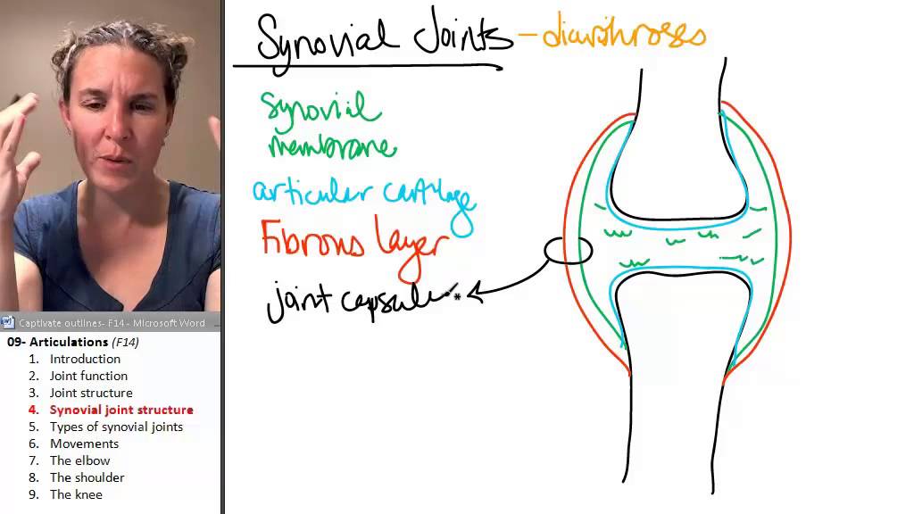
Articulations 4- Synovial joint anatomy
Synovial Joint (Diarthrosis): Definition, Types, Structure ... As the above image shows, there are many types of synovial joint. Starting at (a), the pivot joint can be found in the first few cervical vertebrae, which must twist and turn to allow for rotation of the head and neck. A hinge joint, (b), can be seen in places like the elbow and knee which are only allowed to bend in one direction.
Das Kniegelenk - Anatomie der unteren Extremität
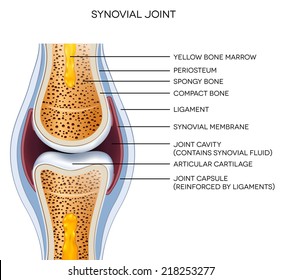
Synovial Joint Diagram Label - schematron.org The structure of a synovial joint is demonstrated by a diagram in which the articulating bones are surrounded by the articular capsule, which comprises an exterior fibrous capsule and an interior synovial membrane. Start studying label the synovial joint. Learn vocabulary, terms, and more with flashcards, games, and other study tools.

Synovial (movable) Joints
TMJ Anatomy - Physiopedia TMJ shown in the box. The temporomandibular joint (TMJ), or jaw joint, is a synovial joint that allows the complex movements necessary for life. It is the joint between condylar head of the mandible and the mandibular fossa of the temporal bone. This system is made up of the TMJ, teeth and soft tissue and it plays a role in breathing, eating ...
Synovial joints Images, Stock Photos & Vectors | Shutterstock
Home Page: Journal of Hand Surgery 28-02-2022 · The cover image shows radiographs of semiconstrained distal radioulnar joint arthroplasty immediately after surgery and 6 years after surgery. Note the collar resorption of the distal ulna. See the article by Brannan et al, “Two-Year Clinical and Radiographic Evaluation of Scheker Prosthesis (Aptis) Distal Radioulnar Joint Arthroplasty” on page 295. .
Solved] Use the list of structures provided to label the ...
Synovial Joint - SmartDraw Synovial Joint. Create healthcare diagrams like this example called Synovial Joint in minutes with SmartDraw. SmartDraw includes 1000s of professional healthcare and anatomy chart templates that you can modify and make your own. 26/37 EXAMPLES.

Human synovial joint | Download Scientific Diagram
Ankle and foot anatomy: Bones, joints, muscles | Kenhub 22-02-2022 · The ankle joint, also known as the talocrural joint, allows dorsiflexion and plantar flexion of the foot. It is made up of three joints: upper ankle joint (tibiotarsal), talocalcaneonavicular, and subtalar joints. The last two together are called the lower ankle joint.

GCSE PE- Synovial Joints structure and Exam question
Joint: synovial - MyDr.com.au A synovial joint is the type of joint found between bones that move against each other, such as the joints of the limbs (e.g. shoulder, hip, elbow and knee). Characteristically it has a joint cavity filled with fluid.

Synovial Joint Stock Illustrations – 640 Synovial Joint Stock ...
Metacarpals: Definition, Location, Anatomy, Function, Diagram The joints between the metacarpal and carpal bones, these are all plane synovial joints, except the thumb as it is a saddle joint (another form of synovial joint) [8]. The five metacarpals form prominent articulations on their base or proximal end, with one or more of the four distal carpal bones [4] :

Describe typical synovial joint with a neat labelled diagram ...
What Is Causing Your Knee Pain? - verywellhealth.com 20-03-2021 · A normal knee joint is surrounded by a membrane, the synovium, which produces a small amount of thick fluid, known as synovial fluid. Synovial fluid helps to nourish the cartilage and keep it slippery. The synovium also has a tough outer layer (the joint capsule) which protects and supports the joint.

A general synovial joint. | Download Scientific Diagram
Structure of a synovial joint pdf - Australia Tutorials ... The main parts of synovial joints are labelled on the synovial joint diagram and described in the table below. A, The egg-shaped ovoid surface represents a characteristic of most synovial joints of the body (e.g., hip joint, radiocarpal joint, knee joint, metacarpophalangeal joint). The diagram shows only the convex member of the joint.
How to describe the composition of the essential structure ...
Anatomy and Physiology Of Synovial Joints - An Overview A joint, which is merged or combined with bones and is departed by a fluid present within the joint cavity are called synovial joints. They are freely movable and the most common type of joints. All limb joints and other joints are examples of synovial joints. Similar to other joints, synovial joints are directly connected to each other with ...
Structure of synovial joint
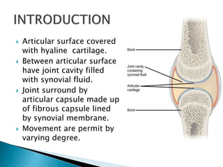
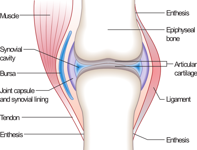
9.4 Synovial Joints - Anatomy & Physiology Figure 9.4.1 - Synovial Joints: Synovial joints allow for smooth movements between the adjacent bones. The joint is surrounded by an articular capsule that defines a joint cavity filled with synovial fluid. The articulating surfaces of the bones are covered by a thin layer of articular cartilage.
Synovial joint activity
Glenoid fossa - Wikipedia Evolution. Interpretations of the fossil remains of Australopithecus africanus (STS 7) and A. afarensis (AL 288-1; a.k.a. Lucy) suggest that the glenoid fossa was oriented more cranially in these species than in modern humans.This reflects the importance of overhead limb postures and suggests a retention of arboreal adaptations in these hominoid primates, whereas the lateral …
Synovial Joint Mechanics | SpringerLink
Types Of Synovial Joints! Trivia Questions Quiz - ProProfs What type of synovial joint is shown in the diagram? Condyloid. Plane. Hinge. Saddle. Ball and Socket. Pivot. The Elbow Joint: Functions And Location! Quiz . The Elbow Joint: Functions And Location! Quiz. How much do you know about the elbow joint, functions, and location? The elbow is a visible joint between the upper and lower parts of the arm.

the generalized structure of a synovial joint Diagram | Quizlet
Solved 3. Label the six different types of synovial joints ... 1.Ball and socket joint: Between humerus and glenoid cavity of scapula 2.Pivot joints between radius …. View the full answer. Transcribed image text: 3. Label the six different types of synovial joints on the diagram by clicking and dragging the labels to the correct location Saddle Joint bok Hinge joint ences 4 Pivot joint Ball and socket N ...
Synovial Joint Structure - TeachPE.com
Hip joint: Bones, movements, muscles | Kenhub Hip joint (Articulatio coxae) The hip joint is a ball and socket type of synovial joint that connects the pelvic girdle to the lower limb. In this joint, the head of the femur articulates with the acetabulum of the pelvic (hip) bone.. The hip joint is a multiaxial joint and permits a wide range of motion; flexion, extension, abduction, adduction, external rotation, internal rotation and ...
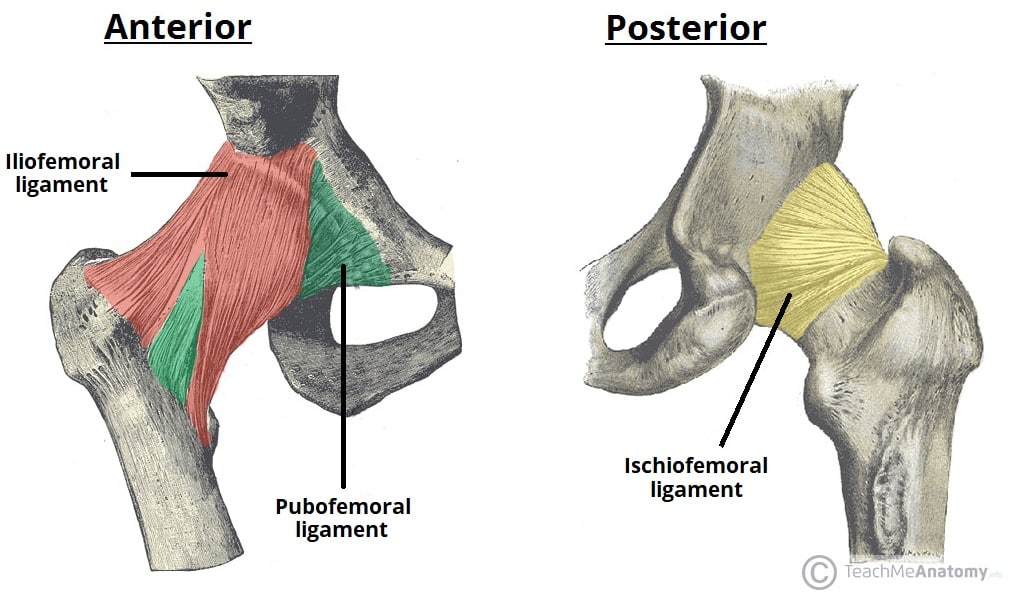
Structures of a Synovial Joint - Capsule - Ligaments ...
Labelled Diagram Of Synovial Joint - schematron.org A synovial joint is a connection between two bones consisting of a cartilage lined As seen in the above picture, the most powerful bite in the world gets its. Synovial joints allow for smooth movements between the adjacent bones. This diagram shows the location of the bursae which are fluid filled sacs in a bone.

Draw labelled diagram Synovial joint. - Biology | Shaalaa.com
Synovial Joint Anatomy in Animal - Definition, Types and ... The synovial joint is a moveable or true joint in an animal's body. Hi there, do you want to learn synovial joint anatomy in animals? Fine, in this article, I will describe the synovial joint structure with a labeled diagram. I will also describe different types of synovial joints in animals. After reading this article, you will know the ...
Synovial joint
Diagram Of Synovial Joint - Elcacerolazo Synovial Joint Diagram Labeled Anatomy Chart With Two Bones Synovial joints are characterized by the presence of a joint cavity. It consists of two layers. Fine in this article I will describe the synovial joint structure with a labeled diagram. Allows movement in only one plane. Tough Fibrous tissue surrounds synovial joints.

Synovial Joints: Structure, Function & Types | Study.com
Synovial joints - Skeletal system - AQA - GCSE Physical ... Synovial joints. A joint is a place where two or more bones meet and is also called an articulation. The role of joints and connective tissue . Connective tissues consist of ligaments, cartilage ...
Structure and function of synovial joints – HSC PDHPE
Structures of a Synovial Joint - Capsule - Ligaments ... A synovial joint is characterised by the presence of a fluid-filled joint cavity contained within a fibrous capsule. It is the most common type of joint found in the human body, and contains several structures which are not seen in fibrous or cartilaginous joints.. In this article we shall look at the anatomy of a synovial joint - the joint capsule, neurovascular structures and clinical ...

All About Joints - Expert how-to for English Riders
Synovial Joints Anatomy Diagram - Quizlet The only 2 synovial joints that aren't diarthrotic. carpals and tarsals. Functional classification of carpals and tarsals. amphiarthrosis. membrane continuous from bone to bone outside the articular capsule. periosteum. fibrous capsule lined by synovial membrane. articular capsule.

Synovial Joint, What is a synovial joint? What is synovial ...
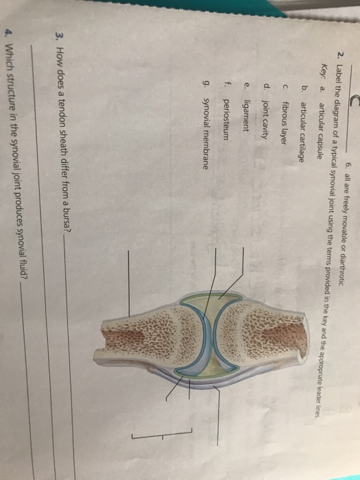
Solved 6. all are freely movable or diarthrotic 2. Label the ...
BTEC Revision Guide Skeletal System

A cross-sectional diagram through a synovial joint. Adapted ...
Diagram of a synovial joint. A synovial joint consists of two ...
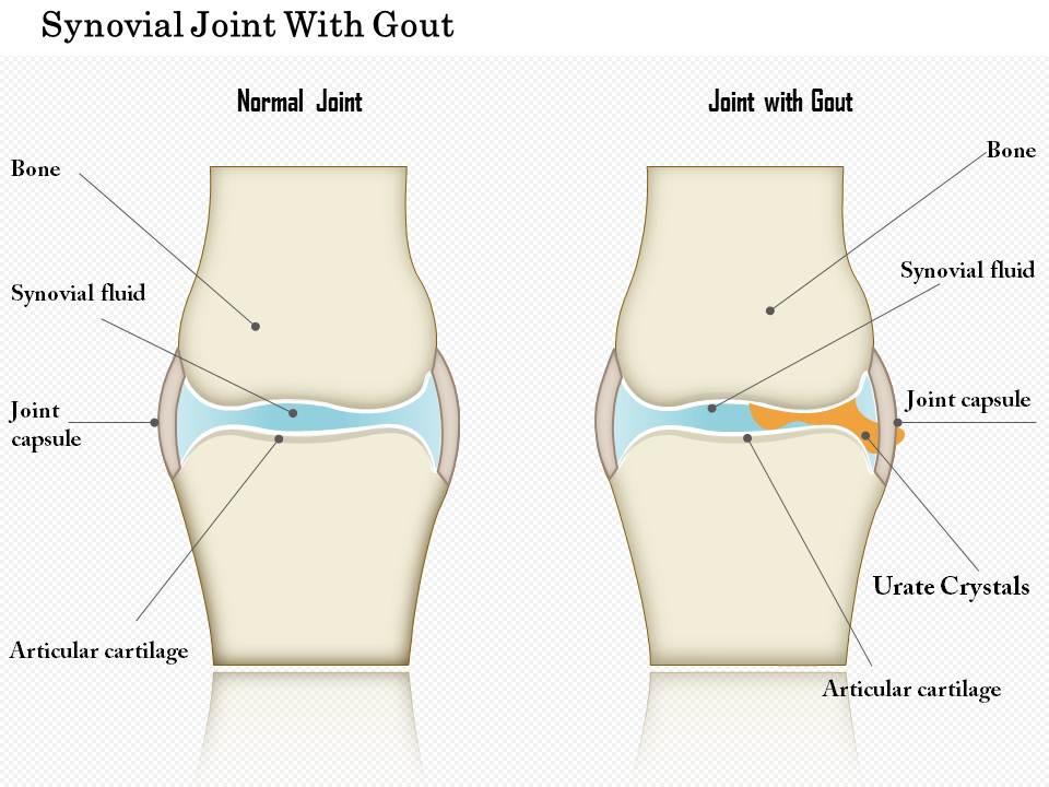
0914 Synovial Joint With Gout Medical Images For PowerPoint ...
Synovial joint - Teaching resources

Synovial Joint Kapselknochen Chart Vektor Abbildung ...

Structure of a synovial joint Diagram | Quizlet
Synovial Joint: Symptoms and Complications

General structure of Synovial Joint Diagram | Quizlet

Synovial Joints: Structure, Function & Types | Study.com
0 Response to "36 diagram of synovial joint"
Post a Comment