36 muscle cell diagram labeled
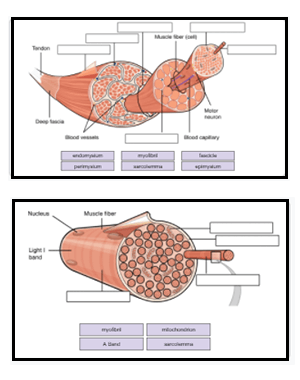
Muscle Cell Structure - Biology MUSCLE CELL STRUCTURE - BIOLOGY. Each muscle fibre is covered by sarcolemma. Tunnel-like extensions pass through the muscle fibre from one side of it to the other in transverse sections through the diameter of the fibre. These tunnel-like extensions are known as transverse tubules. The nuclei of muscle cells are located at the edges of the ... Muscle Cell | Definition, Anatomy, Types & Functions The single muscle cell consists of many nuclei that are pressed against the cell membrane. A muscle cell is a long cell as compared to other kinds of cells, and many muscle cells connect with each other to create the long fibers present in muscle tissue. Muscle Cell Diagram
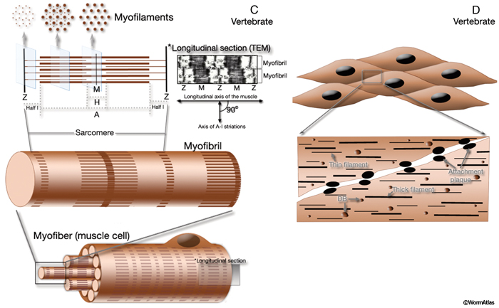
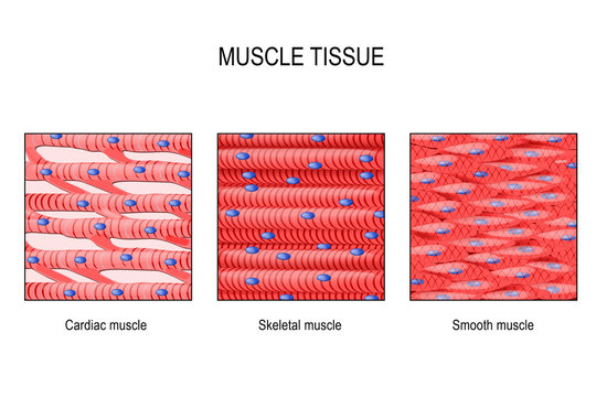
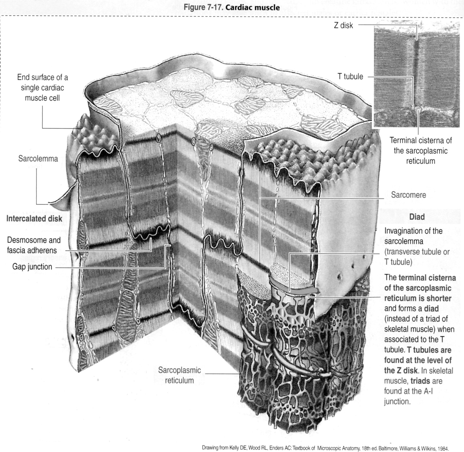
Muscle | histology There are three major types of muscle, and their structure reflects their function. Skeletal and cardiac muscle cells are called striated muscle because of the very regular arrangement of their intracellular contractile units, sarcomeres, at the light microscope (LM) and electron microscope (EM) levels.
Muscle cell diagram labeled
Anatomy, Skeletal Muscle - StatPearls - NCBI Bookshelf The musculoskeletal system comprises one of the major tissue/organ systems in the body. The three main types of muscle tissue are skeletal, cardiac, and smooth muscle groups.[1][2][3] Skeletal muscle attaches to the bone by tendons, and together they produce all the movements of the body. The skeletal muscle fibers are crossed with a regular pattern of fine red and white lines, giving the ... Gram-positive bacteria- cell wall, examples, diseases ... 15.4.2021 · For the gram-positive cell wall, it has a thickness of about 20-80nm thickness made up of a thick peptidoglycan layer outside its cell membrane, unlike the thin layer of gram-negative bacteria (10-15nm) which has a very thin layer of the peptidoglycan of 2-7nm but has a thicker lipid layer making it quite complex than the Gram-positive cell wall. Muscle Cells | Functions & Structure | GCSE Biology Revision Skeletal muscle cells, a barred muscle cell type, it makes the muscle that we use to movement and are categorized into multiple muscle tissues around the entire body, such as that of biceps. Skeletal muscles are connected to bones close to tendons and can be as long as 30 cm, although they are usually 2 to 3 cm in length.
Muscle cell diagram labeled. PDF Muscle Cell Anatomy & Function Human Anatomy & Physiology: Muscle Physiology; Ziser Lecture Notes, 2006 1 Muscle Cell Anatomy & Function (mainly striated muscle tissue) General Structure of Muscle Cells (skeletal) several nuclei (skeletal muscle) skeletal muscles are formed when embryonic cells fuse together some of these embryonic cells remain in the adult and can replace Skeletal Muscle Histology Slide Identification and Labeled ... The skeletal muscle fibers are elongated, cylindrical and multinucleated cells whose length may vary in different animals. In this short guide, you will get a basic concept of skeletal muscle histology from the real slide and labeled diagram. You will also get the identification points of skeletal muscle histology slide with a little description here in this guide. Muscle cell - Wikipedia Structure. The unusual microscopic anatomy of a muscle cell gave rise to its own terminology. The cytoplasm in a muscle cell is termed the sarcoplasm; the smooth endoplasmic reticulum of a muscle cell is termed the sarcoplasmic reticulum; and the cell membrane in a muscle cell is termed the sarcolemma. The sarcolemma receives and conducts stimuli. Skeletal muscle cells Muscles Notes: Diagrams & Illustrations | Osmosis NOTES NOTES MUSCLES MUSCULAR SYSTEM ANATOMY & PHYSIOLOGY osms.it/muscle-anatomy-physiology Three types of muscle cell/tissue Skeletal, cardiac, smooth Differ in location, innervation, cell structure All cells excitable, extensible, elastic SKELETAL MUSCLE Attaches to bone/skin; mostly voluntary; maintains posture, stabilizes joints, generates heat Most muscles consist of belly (contracts ...
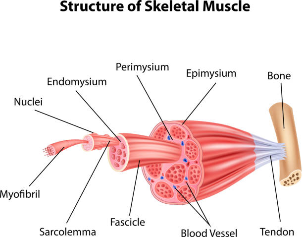
Muscular System - Muscles of the Human Body Skeletal muscle fibers differ dramatically from other tissues of the body due to their highly specialized functions. Many of the organelles that make up muscle fibers are unique to this type of cell. The sarcolemma is the cell membrane of muscle fibers. The sarcolemma acts as a conductor for electrochemical signals that stimulate muscle cells. Types of muscle cells: Characteristics, location, roles ... Muscle cells, commonly known as myocytes, are the cells that make up muscle tissue. There are 3 types of muscle cells in the human body; cardiac, skeletal, and smooth. Skeletal muscle cells are long, cylindrical, multi-nucleated and striated . Each nucleus regulates the metabolic requirements of the sarcoplasm around it. Structure of Skeletal Muscle | SEER Training Structure of Skeletal Muscle. A whole skeletal muscle is considered an organ of the muscular system.Each organ or muscle consists of skeletal muscle tissue, connective tissue, nerve tissue, and blood or vascular tissue.. Skeletal muscles vary considerably in size, shape, and arrangement of fibers. They range from extremely tiny strands such as the stapedium muscle of the middle ear to large ... 10.2 Skeletal Muscle - Anatomy & Physiology Figure 10.2.2 - Muscle Fiber: A skeletal muscle fiber is surrounded by a plasma membrane called the sarcolemma, which contains sarcoplasm, the cytoplasm of muscle cells. A muscle fiber is composed of many myofibrils, which contain sarcomeres with light and dark regions that give the cell its striated appearance.
Label the muscle cell diagram Quiz - PurposeGames.com This is an online quiz called Label the muscle cell diagram . There is a printable worksheet available for download here so you can take the quiz with pen and paper. Your Skills & Rank. Total Points. 0. Get started! Today's Rank--0. Today 's Points. One of us! Game Points. 16. Cardiomyocytes (Cardiac Muscle Cells)- Structure, Function ... Although the regeneration of cardiac muscle cells was thought to be absent, studies have shown that these cells renew at a significantly low rate throughout the life of an individual. For instance, for younger people, about 25 years of age, the annual turnover of cardiomyocytes is said to be about 1 percent. Learn all muscles with quizzes and labeled diagrams | Kenhub Human body muscle diagrams. Muscle diagrams are a great way to get an overview of all of the muscles within a body region. Studying these is an ideal first step before moving onto the more advanced practices of muscle labeling and quizzes. If you're looking for a speedy way to learn muscle anatomy, look no further than our anatomy crash courses . Label structure of skeletal muscle Diagram | Quizlet Label structure of skeletal muscle. STUDY. Learn. Flashcards. Write. Spell. Test. PLAY. Match. Gravity. Created by. Erica_Hartfield. Terms in this set (10) myofibrils. a long, filamentous organelle found within muscle cells that has a banded appearance. tendon. cordlike extension of connective tissue beyond the muscle, serving to attach it to ...
How Many Muscles Are in the Human Body? Plus a Diagram
Labelled diagram of a muscle cell | Human anatomy model ... Our musculoskeletal system is made up of muscles, tendons, ligaments, bones, cartilage, joints and bursae. Our muscles work with the nervous system to contract when stimulated with impulses from motor nerves. Little Colleen. CCMT. Human Body Anatomy. Muscle Anatomy. Eye Anatomy Diagram. Eyeball Anatomy.
Which cells consist of more than 1 nucleus? - Quora
Muscle Cell (Myocyte): Definition, Function & Structure ... Muscle Cell Definition. A muscle cell, known technically as a myocyte, is a specialized animal cell which can shorten its length using a series of motor proteins specially arranged within the cell. While several associated proteins help, actin and myosin form thick and thin filaments which slide past each other to contract small units of a muscle cell. . These units are called sarcomeres, and ...
3,206 Muscle Cell Stock Photos, Pictures & Royalty-Free ...
Magnesium sensing via LFA-1 regulates CD8+ T cell effector ... 19.1.2022 · The T cell co-stimulatory molecule LFA-1 binds extracellular magnesium ions, allowing it to modulate murine and human CD8+ T cell effector function in response to magnesium levels during normal responses and in the context of immunotherapy.

The basic morphology of the cardiac muscle cell. Top: The ...
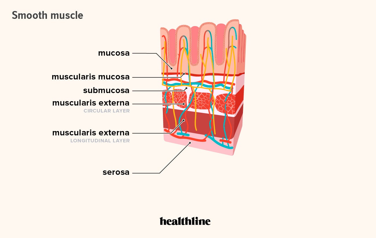
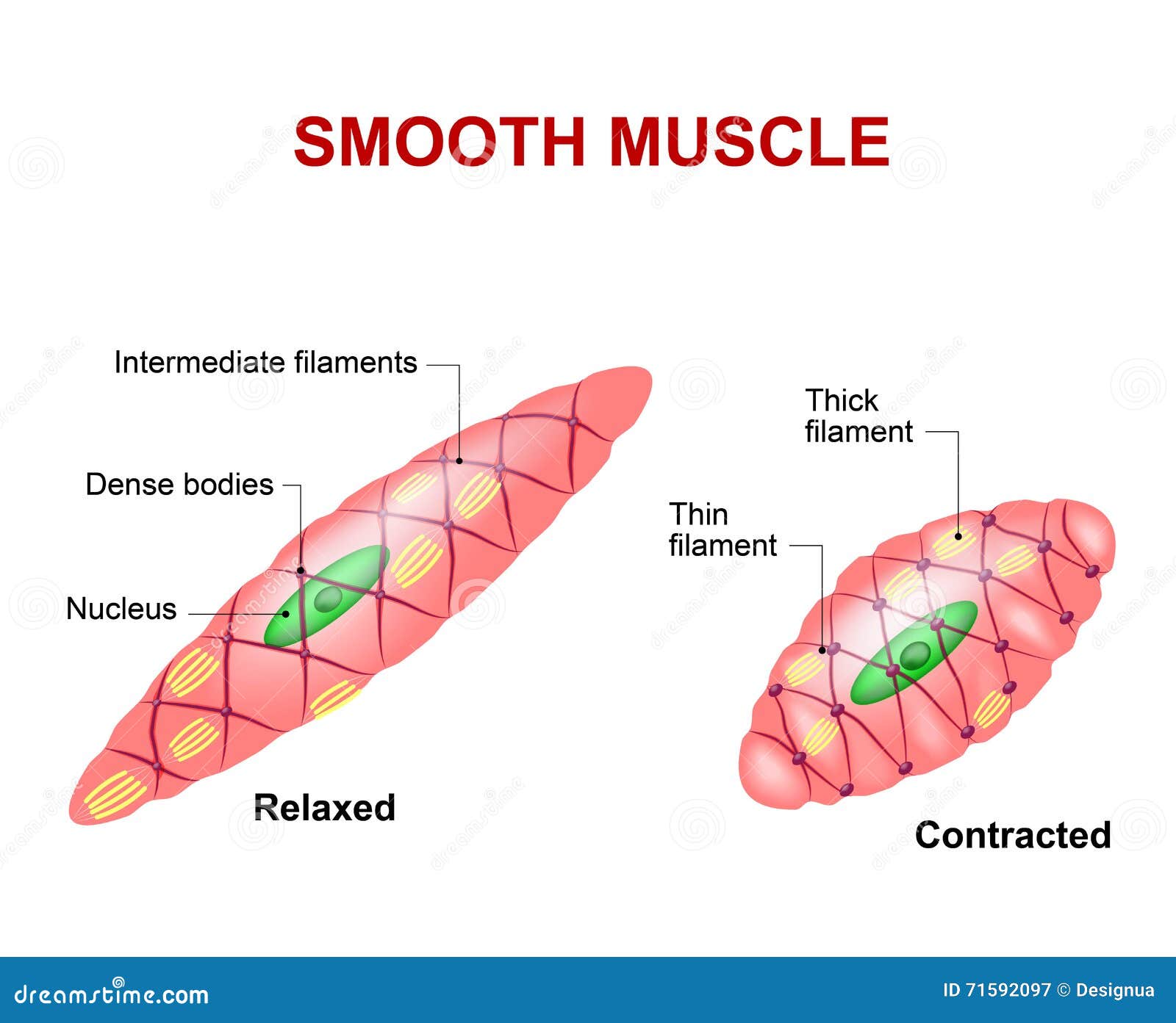
Smooth muscle: Structure, function, location | Kenhub Structure. The smooth muscle cell is 3-10 µm thick and 20-200 µm long. The cytoplasm is homogeneously eosinophilic and consists mainly of myofilaments. The nucleus is located in the center and takes a cigar-like shape during contraction. The cell membrane forms small pouch-like invaginations into the cytoplasm (caveolae) which are functionally equivalent to the T-tubules of the skeletal ...
Microanatomy of Muscles
Muscle Cells | Functions & Structure | GCSE Biology Revision Skeletal muscle cells, a barred muscle cell type, it makes the muscle that we use to movement and are categorized into multiple muscle tissues around the entire body, such as that of biceps. Skeletal muscles are connected to bones close to tendons and can be as long as 30 cm, although they are usually 2 to 3 cm in length.

Draw a diagram of muscle Label on it (i) nucleus (ii) spindle ...
Gram-positive bacteria- cell wall, examples, diseases ... 15.4.2021 · For the gram-positive cell wall, it has a thickness of about 20-80nm thickness made up of a thick peptidoglycan layer outside its cell membrane, unlike the thin layer of gram-negative bacteria (10-15nm) which has a very thin layer of the peptidoglycan of 2-7nm but has a thicker lipid layer making it quite complex than the Gram-positive cell wall.
Handbook - Muscle System Introduction
Anatomy, Skeletal Muscle - StatPearls - NCBI Bookshelf The musculoskeletal system comprises one of the major tissue/organ systems in the body. The three main types of muscle tissue are skeletal, cardiac, and smooth muscle groups.[1][2][3] Skeletal muscle attaches to the bone by tendons, and together they produce all the movements of the body. The skeletal muscle fibers are crossed with a regular pattern of fine red and white lines, giving the ...

Neuron - Wikipedia

Skeletal Muscle Physiology

Muscle Cell, Illustration - Stock Image - C043/2741 - Science ...
Muscle cell - Wikipedia

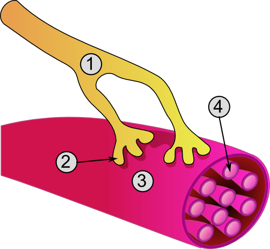
Clip Art Vector - Neuromuscular junction vector illustration ...
Cardiac Muscle Images – Browse 13,691 Stock Photos, Vectors ...

File:Diagram of muscle cells CRUK 035.svg - Wikimedia Commons

Draw a labeled diagram of smooth muscle.

Skeletal Muscle Cell Diagram | Quizlet

Copyright © The McGraw-Hill Companies. Chapter 10. Muscle ...
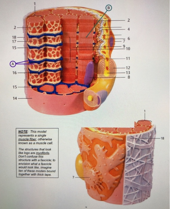
Solved B. Examine the three-dimensional model of a skeletal ...

Ch. 10 - Types of muscle tissue Diagram | Quizlet

Chapter 12 | Muscle diagram, Smooth muscle tissue, Cell diagram

Immunoperoxidase labelling of smooth muscle cells surrounding ...
Microanatomy of Muscles
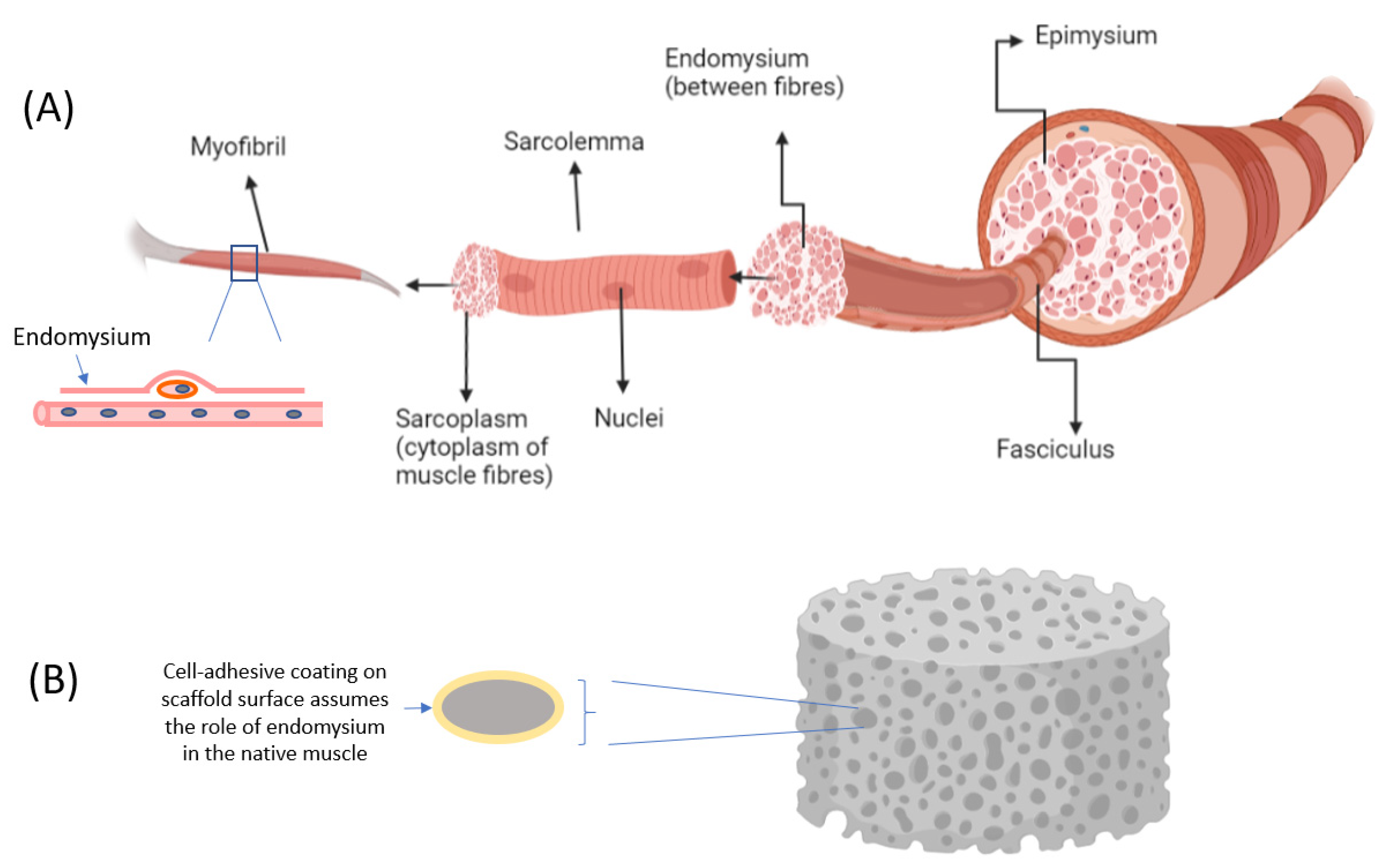
Micromachines | Free Full-Text | Systems for Muscle Cell ...
Muscle Cell Stock Illustrations – 4,064 Muscle Cell Stock ...

Smooth Muscle Anatomy for Biology Science Education Stock ...
Muscles Labeling

10.7 Smooth Muscle Tissue – Anatomy & Physiology

Muscle Cell labeling Diagram | Quizlet

Muscle fiber structure and inner parts anatomical description ...

FACS isolation of GFP-labeled body wall muscle cells. A ...
Histology Laboratory Manual

labelled diagram - Aaiza site

10.2 Skeletal Muscle – Anatomy & Physiology

Labelled diagram of a muscle cell | Muscle, Muscle structure ...

Draw a diagram of muscle. Label on it (i) nucleus (ii ...
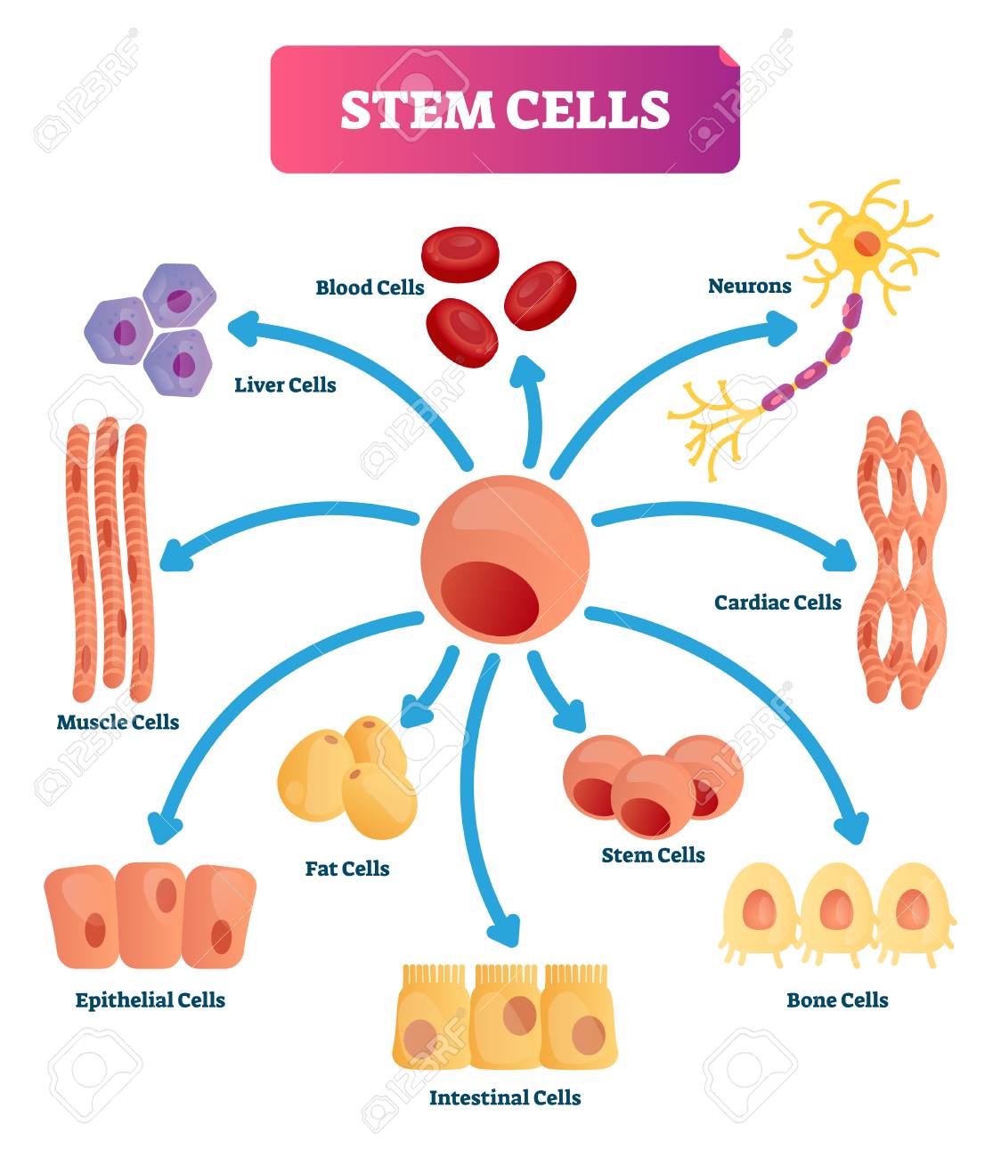
Stem Cells Vector Illustration. Medical Labeled Diagram With ...
0 Response to "36 muscle cell diagram labeled"
Post a Comment