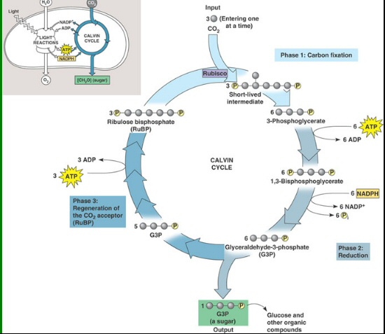
40 drag the labels to their appropriate locations on the cycle diagram below.
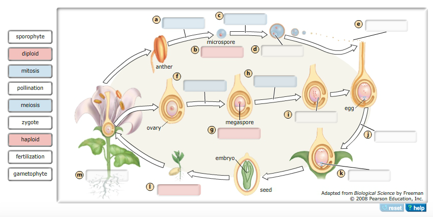
[Expert Answer] Instructions: Drag each label to the ... Instructions: Drag each label to the correct location on the image. Identify each passage as either functional text or expository text. Link to assignment photo below Part a alternation of generations in ... - Course Hero Drag the labels to their appropriate locations on the diagram of the angiosperm life cycle. Labels can be used once or more than once. Use only white labels for white targets, pink labels for pink targets, and blue labels for blue targets. True False You did not open hints for this part.
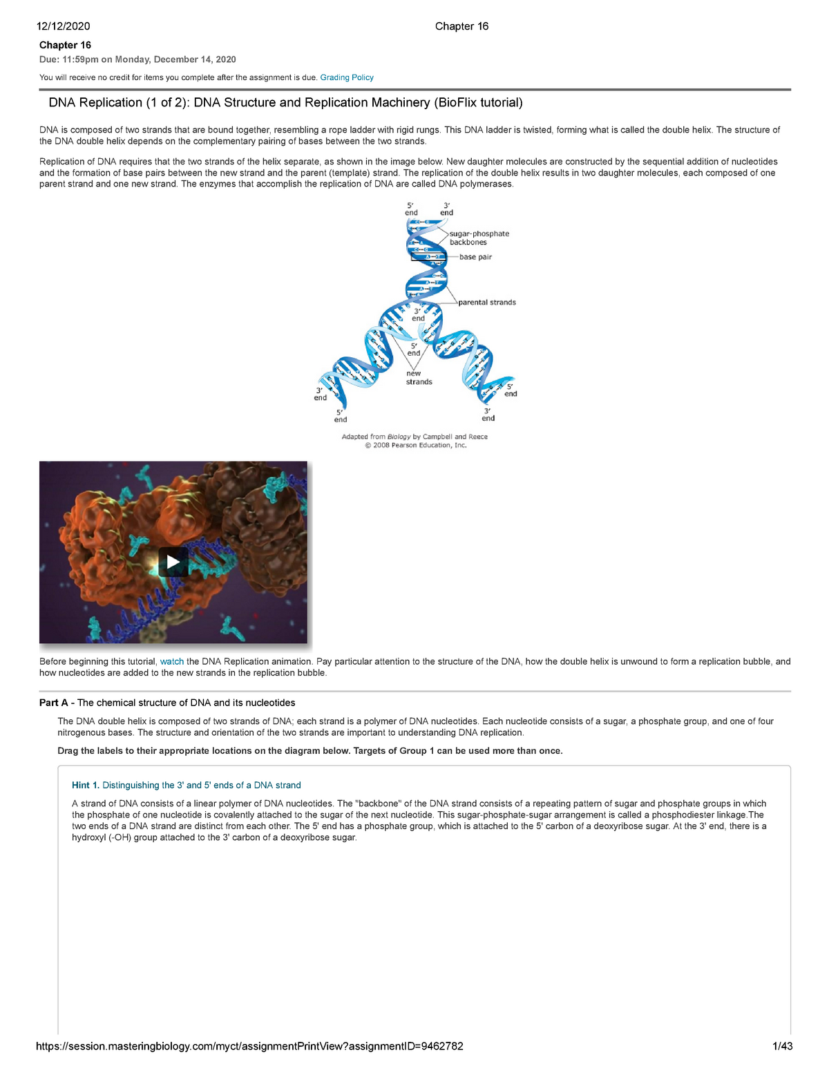
Mastering Biology Chapter 16 - RHS Homework Drag the labels to their appropriate locations on the diagram below. Pink labels can be used more than once. ... The diagram below shows a replication bubble with synthesis of the leading and lagging strands on both sides of the bubble. The parental DNA is shown in dark blue, the newly synthesized DNA is light blue, and the RNA primers ...

Drag the labels to their appropriate locations on the cycle diagram below.
Mastering Biology Chp. 13 HW - Subjecto.com Drag the labels to their appropriate locations on the diagram below. Pink labels can be used more than once. [The DNA double helix is constructed from two strands of DNA, each with a sugar-phosphate backbone and nitrogenous bases that form hydrogen bonds, holding the two strands together. Each DNA strand has two unique ends. Mastering Biology Chp. 10 HW - Subjecto.com Complete the diagram to show the life cycle of a typical animal. Follow these steps: 1. First, drag blue labels onto blue targets only to identify each stage of the life cycle. 2. Next, drag pink labels onto pink targets only to identify the process by which each stage occurs. 3. Diagram Of Brain with their Labelings and Detailed Explanation The forebrain is the anterior part of the brain, which comprises the cerebral hemispheres, the thalamus, and the hypothalamus. It also consists of two subdivisions called the telencephalon and diencephalon. Along with the optic nerves and cranial nerves, the forebrain also includes the olfactory system, or sense of smell as well as the lateral ...
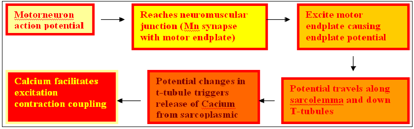
Drag the labels to their appropriate locations on the cycle diagram below.. Which of these provides the source for poetry, according ... Which of the following could be a sequence in the carbon cycle? ... . this concentration gradient is key to the movement of ca2+ in the muscle cell in response to an action potential. the cycle diagram below shows the sequence of events that affect ca2+ levels in a muscle cell, beginning with the propagation of an action potential down a t ... Part C - How an action potential affects Ca2+ movement in ... The cycle diagram below shows the sequence of events that affect Ca2+ levels in a muscle cell, beginning with the propagation of an action potential down a T tubule (top of the diagram). Drag the labels to their appropriate locations on the cycle diagram below. Note that SR stands for sarcoplasmic reticulum. • Show less Drag the correct label to each event. Each label can be ... Drag each label to the correct location. The labels can be used more than once. Match each statement with the type of weathering it describes. chemical weathering or Mechanical Weathering. 1. Weathering is caused by precipitation reacting with minerals in the rock.- Chemical Weathering. 2. Weathering is caused by freezing and thawing of water.- The diagram below illustrates the life cycle of... ask 8 The diagram below illustrates the life cycle of Dictyostelium, a cellular slime mold. Label the diagram to indicate the processes that are occurring as well as whether each cell type is haploid or diploid. Drag the labels to their appropriate locations on the diagram. Use the pink labels for pink targets.
Ch 38 HW Flashcards - Quizlet Label structures and processes (using white labels), indicate whether different structures are haploid or diploid (using pink labels), and indicate the types of cell division that occur at different points in the life cycle (using blue labels). Drag the labels to their appropriate locations on the diagram of the angiosperm life cycle. Which of the following is not true concerning the usage of ... All layers of the Earth below the crust are liquid. ... . this concentration gradient is key to the movement of ca2+ in the muscle cell in response to an action potential. the cycle diagram below shows the sequence of events that affect ca2+ levels in a muscle cell, beginning with the propagation of an action potential down a t tubule (top of ... Assignment 14 Flashcards - Quizlet The cycle diagram below shows the sequence of events that affect Ca2+ levels in a muscle cell, beginning with the propagation of an action potential down a T tubule (top of the diagram). Drag the labels to their appropriate locations on the cycle diagram below. Note that SR stands for sarcoplasmic reticulum. Indicate whether each description applies to a specific ... Indicate which structures in the life cycle are haploid or diploid (using labels of Group 2), and label the processes (using labels of Group 1) and stages (using labels of Group 3). Drag the labels to their appropriate locations on the diagram. Labels may be used more than once.
Chpt. 1, 3, 24, and 9 HW - Weebly Drag the appropriate labels to their respective targets. (left from top to bottom) s, g1, cytokinesis (right from top to bottom) interphase, g2, mitosis, mitotic phase 14. During which phase of the cell cycle does DNA duplication, or replication, take place? Interphase When a double helix of DNA is replicated, two complete helices are formed. Drag the labels from the left to their correct locations First, drag blue labels onto blue targets only to identify each stage of the life cycle. Next, drag pink labels onto pink targets only to identify the process by which each stage occurs. Then, drag white labels onto white targets only to identify the ploidy level at each stage. Labels can be used once, more than once, or not at all. Solved Life cycle of a gymnosperm The diagram below ... Life cycle of a gymnosperm The diagram below illustrates the life cycle of one type of gymnosperm, a conifer. Indicate which structures in the life cycle are haploid or diploid (using pink labels), and label the processes (using blue labels) and stages (using white labels). Drag the labels to their appropriate locations on the diagram. › 1814485 › Taiz_and_Zeiger_Plant(PDF) Taiz & Zeiger- Plant Physiology | Munish K Bansal ... Academia.edu is a platform for academics to share research papers.
Mitosis vs. Meiosis: Key Differences, Chart and Venn Diagram ...
Part A Hydrogen bonding Label the following diagram of ... Part A - Hydrogen bonding Label the following diagram of water molecules, indicating the location of bonds and the partial charges on the atoms. Drag the labels to their appropriate locations on the diagram of the water molecules below. Labels can be used once, more than once, or not at all. Hint 1.

Neoantigens in cancer immunotherapy
Diagram of the Rock Cycle Explained - Rock and Mineral Planet Diagram of the Rock Cycle Explained. A diagram of the rock cycle is a way to explain the formation, or deformation, of the three types of rocks we find on our earth; sedimentary, metamorphic, and igneous. The rock cycle picture diagram shows an upper level view of the process in which these types of rocks will be created or transformed.

Solved Drag the labels from the left to their correct | Chegg.com
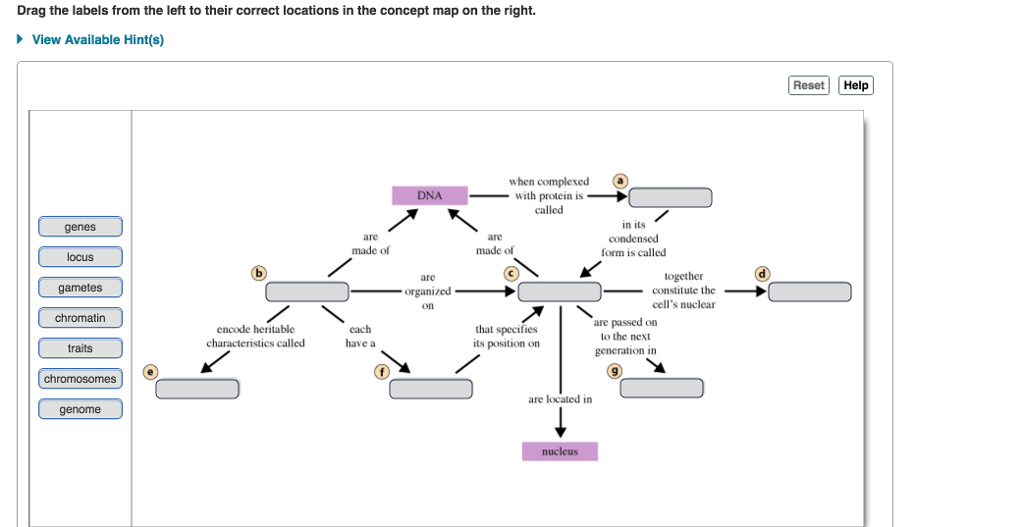
Drag Each Image To The Phase Of Meiosis I It Depicts Drag the labels onto the diagram to identify the stages of the cell cycle. Drag the blue labels to the blue targets to identify the stage of. A cell with a diploid number of 20 undergoes meiosis. Drag the labels from the left to their correct locations in the concept map on the right. Here in these phase the chromosome are.
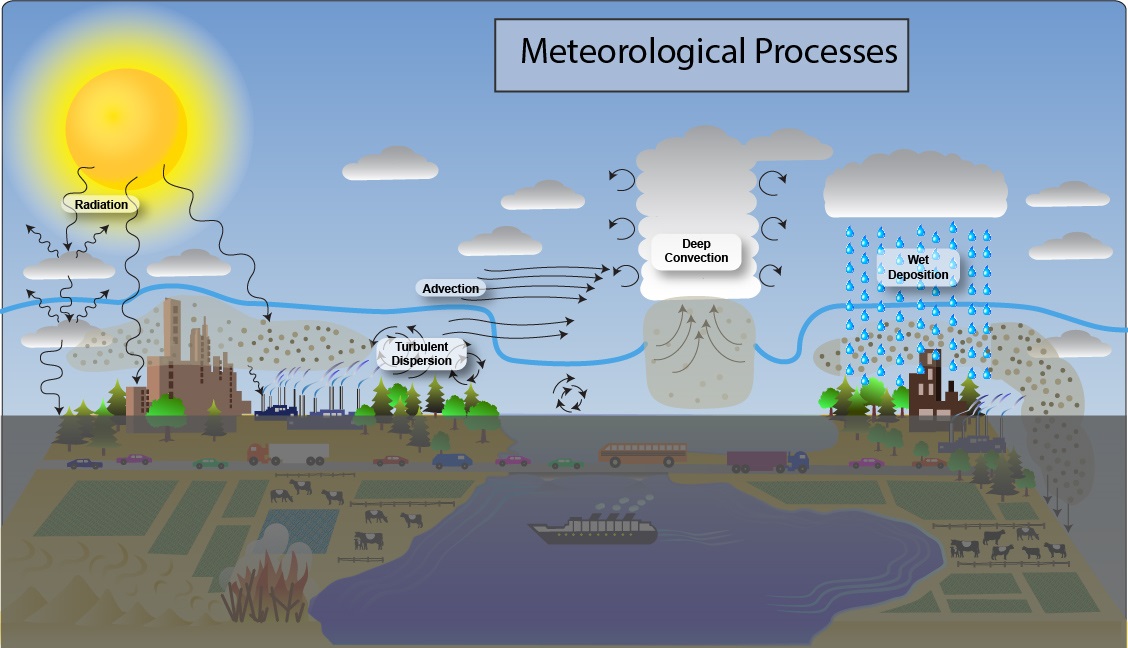
Meteorological Process Overview | US EPA
Art-labeling Activities Correctly match the terms with the correct location of the diagram of red blood cell turnover. For each item below, use the pull-down menu to select the letter that labels the correct part of the image. 1.1 biliverdin: 1.2 bilirubin: 1.3 heme: 1.4 hemolysis ... Match the terms in their appropriate locations in the diagram of the coagulation ...
A First Reconnaissance of the Atmospheres of Terrestrial ...
(Get Answer) - The diagram below shows a length of DNA ... The diagram below shows a length of DNA containing a bacterial gene. Drag the labels to their appropriate locations in the diagram to describe the function or characteristics of each part of the gene. Not all labels will be use.
Working with Molecular Genetics Chapter 5, DNA Replication I ...
Solved Drag the labels to their appropriate locations on ... Transcribed image text: Drag the labels to their appropriate locations on the cycle diagram below. Note that SR stands for sarcoplasmic reticulum. cytosolic Ca2+ level rises Ca2+ diffuses out of myofibril cytosolic Ca2+ level drops Ca2+ diffuses into myofibril Ca2+ diffuses out of SR Ca2+ pumped into SR action potential propagates down t tubule Ca2+ channels in SR open Ca2+ channels in SR ...
Flexible Manufacturing Systems Optimization with Meta ...
Alternation Of Generations Life Cycle Diagram The diagram below illustrates the alternation of generations that is characteristic of the angiosperm life cycle. Label structures and processes (using white labels), indicate whether different structures are haploid or diploid (using pink labels), and indicate the types of cell division that occur at different points in the life cycle (using blue labels).
Unit 3
› 18101048 › Romer_Advanced(PDF) Romer Advanced Macroeconomics | Jose Sousa - Academia.edu Academia.edu is a platform for academics to share research papers.
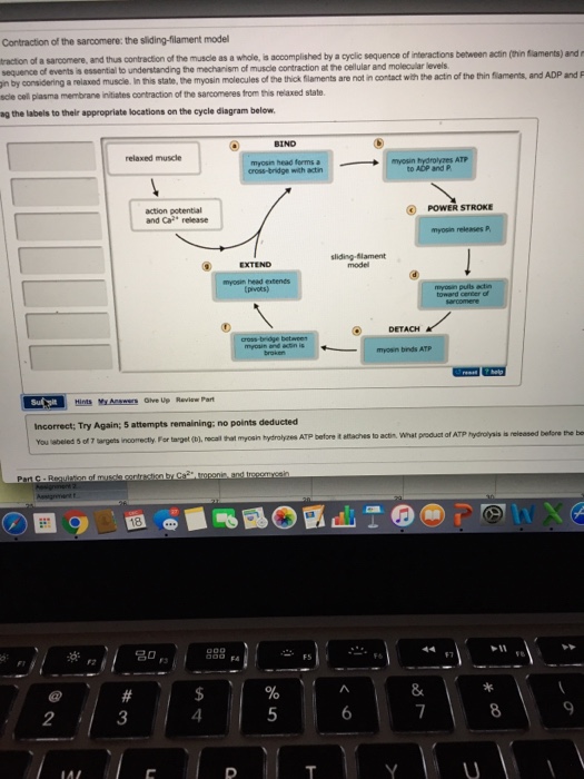
Solved Contraction of the sarcomere the sliding filament ...
Mastering Biology: Chapter 7 Flashcard Example #6180 ... Drag the labels to their appropriate locations in the figure. First, drag labels to targets (a) and (b) to indicate whether these environments are hydrophilic or hydrophobic. Next, drag the phospholipid layers to targets (c) and (d) to indicate how they are oriented in the plasma membrane.

Drag and drop Question Types - Powered by Kayako Help Desk ...
44 place the correct labels on the appropriate area of the ... Drag the correct labels to the appropriate locations in the diagram to show the composition of the daughter duplexes after one and two cycles of dna replication. Assign the appropriate labels to the phase diagram shown below. 1st attempt see periodic t dep. Solved 10 Question 1 Point Place The...
Mastering Biology Chapter 15 – RHS Homework
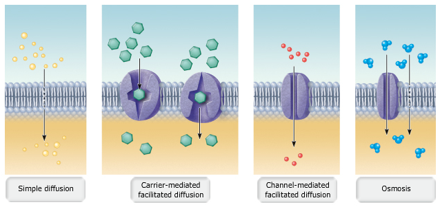
Answered: 18. Label the following diagram: Part A… | bartleby 18. Label the following diagram: Part A - Diffusion Drag the labels to their appropriate locations on the diagram. Side with higher concentration of molecules (b) Plasma membrane Side with lower concentration of molecules Diffusion causes a net movement of molecules down their concentration gradient.

F-1 1 a2242683zf-1.htm F-1 Use these links to rapidly review ...
33 Label The Following Diagram Of ... - Labels For Your Ideas Drag the labels to their appropriate locations on the diagram of the water molecules below. Label the following diagram of water molecules indicating the location of bonds and the partial charges on the atoms. There are two lone pairs of electrons on each oxygen atom represented by. Labels can be used once more than once or not at all.
Mastering biololgy-Chapter 16 - BIO 210 - Biology I - BMCC ...
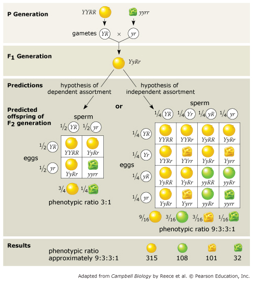
Mastering Biology Chapter 15 - RHS Homework Drag the labels to their appropriate locations to complete the Punnett square for Morgan's F1x F1cross. Drag pink labels onto the pink targets to indicate the alleles carried by the gametes (sperm and egg). Drag blue labels onto the blue targets to indicate the possible genotypes of the offspring.

Fertilization and seed developement.docx - Fertilization and ...
Diagram Of Brain with their Labelings and Detailed Explanation The forebrain is the anterior part of the brain, which comprises the cerebral hemispheres, the thalamus, and the hypothalamus. It also consists of two subdivisions called the telencephalon and diencephalon. Along with the optic nerves and cranial nerves, the forebrain also includes the olfactory system, or sense of smell as well as the lateral ...
Drag and drop onto image question type - MoodleDocs
Mastering Biology Chp. 10 HW - Subjecto.com Complete the diagram to show the life cycle of a typical animal. Follow these steps: 1. First, drag blue labels onto blue targets only to identify each stage of the life cycle. 2. Next, drag pink labels onto pink targets only to identify the process by which each stage occurs. 3.
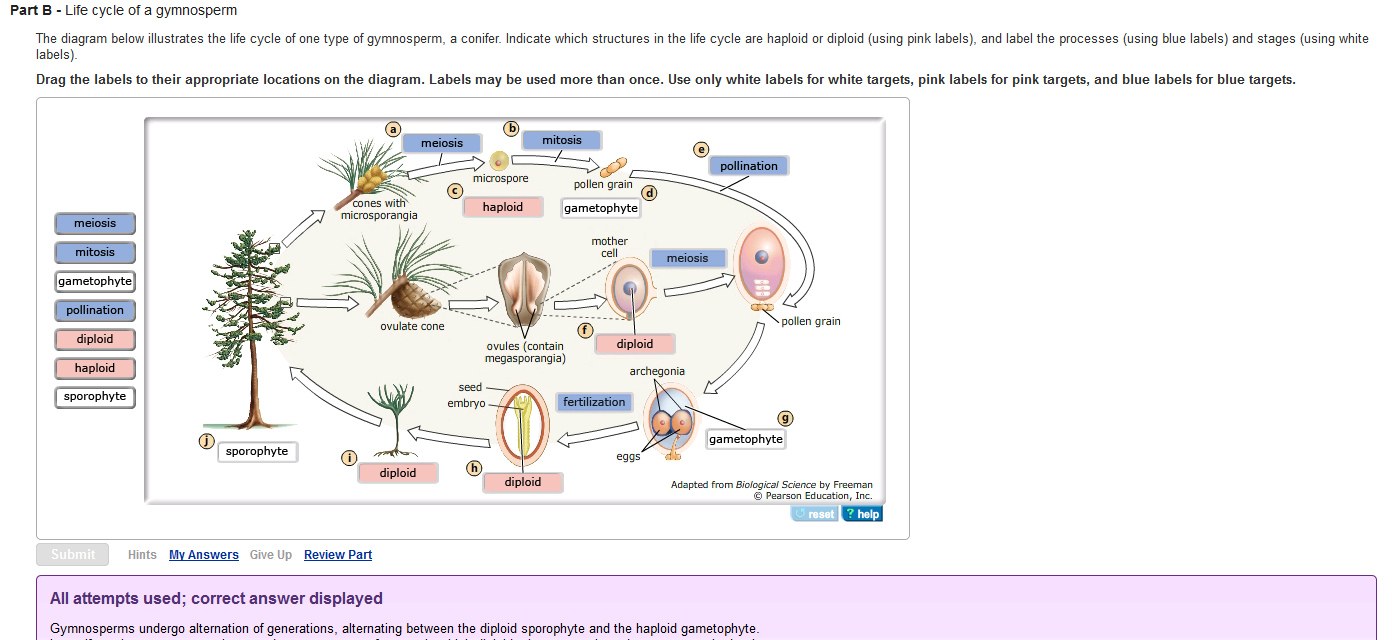
Solved Life cycle of a gymnosperm The diagram below | Chegg.com
Mastering Biology Chp. 13 HW - Subjecto.com Drag the labels to their appropriate locations on the diagram below. Pink labels can be used more than once. [The DNA double helix is constructed from two strands of DNA, each with a sugar-phosphate backbone and nitrogenous bases that form hydrogen bonds, holding the two strands together. Each DNA strand has two unique ends.

ANATOMY AND PHYSIOLOGY - ScienceDirect

Drag and drop Question Types - Powered by Kayako Help Desk ...
Twinkle Toes Engineering
Issue boards | GitLab

Chapter 11 Homework Flashcards | Quizlet

Chapter 9 Homework Flashcards | Quizlet
Solved Drag the labels from the left to their correct | Chegg.com
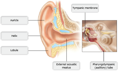
A&P Chapter 15 The Special Senses Flashcards - Easy Notecards

Krebs Cycle - Biology
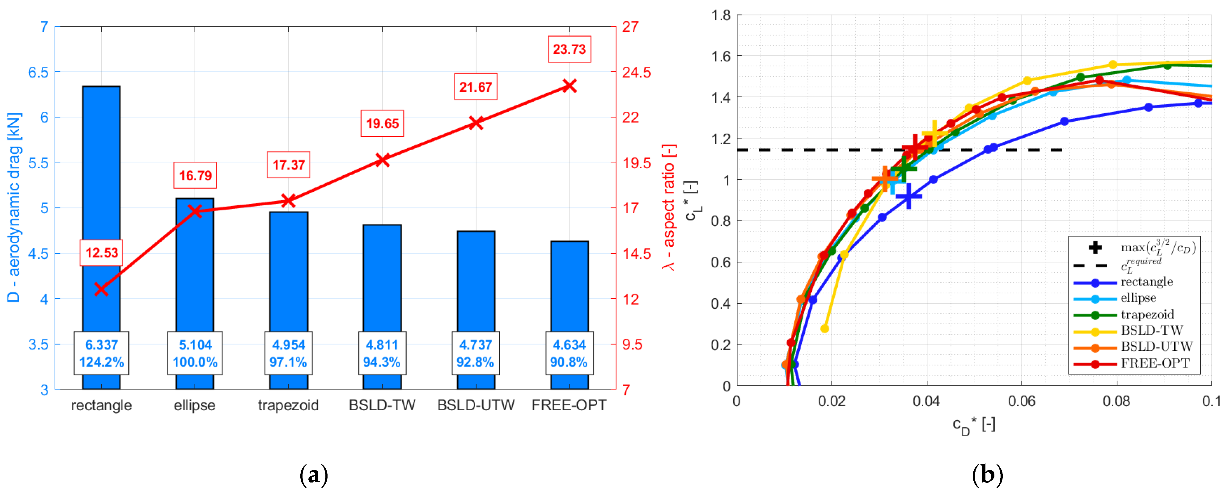
Aerospace | Free Full-Text | Aerodynamic Design and Strength ...

EXAM 2 Cell Biology (Mastering) Flashcards | Quizlet
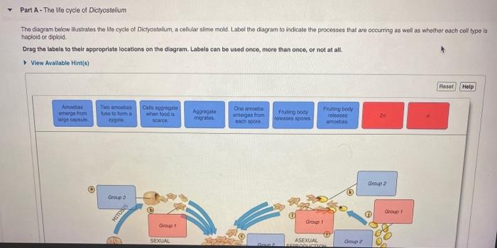
Solved Part A - The We cycle of Dictyostelium The diagram ...
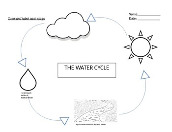
Water Cycle Label Teaching Resources | Teachers Pay Teachers
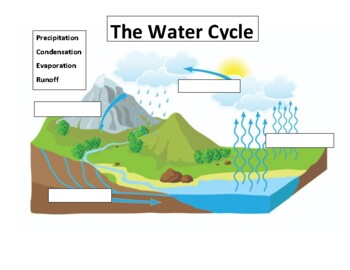
Water Cycle Label Teaching Resources | Teachers Pay Teachers

Drag each label to the correct location on the diagram. Each ...

Chapter 9 Homework Flashcards | Quizlet
Novel Regulators in Photosynthetic Redox Control of Plant ...
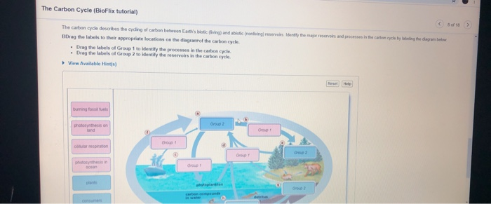
Solved The Carbon Cycle (BioFlix tutorial) the d e cade The ...
Biological Molecules
Medical Management Of Adults With Neurologic Disabilities
144476606-Gene-Regulation
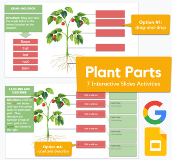
Plant Parts Labels Teaching Resources | Teachers Pay Teachers
A&P Chapter 3 Cells: The Living Units Flashcards - Easy Notecards
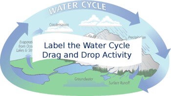
Water Cycle Label Teaching Resources | Teachers Pay Teachers
0 Response to "40 drag the labels to their appropriate locations on the cycle diagram below."
Post a Comment