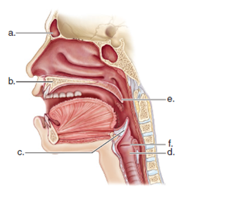
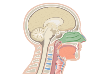
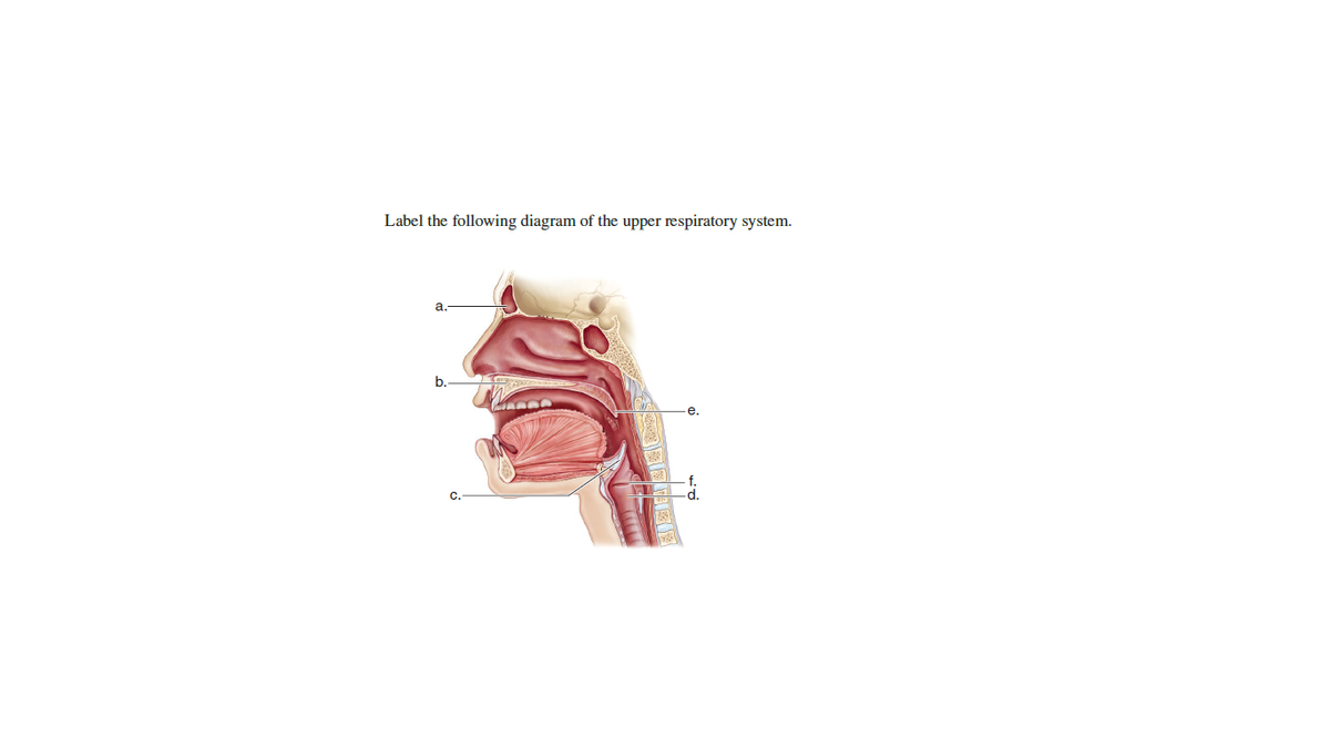
40 complete the labeling of the diagram of the upper respiratory
Labeled Sagittal Section of the Upper Respiratory ... Start studying Labeled Sagittal Section of the Upper Respiratory Structures. Learn vocabulary, terms, and more with flashcards, games, and other study tools. Resp review - NAME _ LAB TIME/DATE Anatomy ... - Course Hero LAB TIME/DATE NAME _ Anatomy ofthe Respiratory System Upper and Lower RespiratorySystem Structures 1. Complete the labeling of the diagram of the upper respiratory structures (sagittal section).
BI217 Sec.750 Spring 2017: Exercise 36 Flashcards | Quizlet Complete the labeling of the diagram of the upper respiratory structures (sagittal section). 2. Two pairs of vocal folds are found in the larynx. Which pair are the true vocal cords (superior or inferior)? Inferior 3. Name the specific cartilages in the larynx that correspond to the following descriptions. A.) forms the Adam's apple:

Complete the labeling of the diagram of the upper respiratory
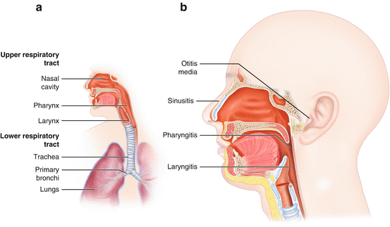
Exercise 36 Review Sheet Anatomy of the Respiratory System ... Exercise 36 Review Sheet Anatomy of the Respiratory System Name Lab Time/DateUpper and Lower Respiratory System Structures 1 Complete the labeling of the diagram of the upper respiratory structures (sagittal section). 2 Two pairs of vocal folds are found in the larynx. Solved: Chapter E23 Problem 1E Solution | Laboratory ... Step 1 of 3 The respiratory tract is divided into two parts. They are upper respiratory tract and lower respiratory tract. Upper respiratory tract includes various passages and structures such as nose, nasal cavity, mouth, throat, and larynx (voice box). The entire respiratory tract is lined with a mucous membrane that secretes mucus. Upper Respiratory System (Labeling) Diagram | Quizlet Start studying Upper Respiratory System (Labeling). Learn vocabulary, terms, and more with flashcards, games, and other study tools.
Complete the labeling of the diagram of the upper respiratory. Upper Respiratory Tract Labeling Diagram | Quizlet Start studying Upper Respiratory Tract Labeling. Learn vocabulary, terms, and more with flashcards, games, and other study tools. PDF PC\|MAC Created Date: 4/28/2015 1:20:29 PM Respiratory System Labelling Worksheet Image Result For ... 31 Complete The Labeling Of The Diagram Of The Upper Respiratory ... Lung worksheetLungs Worksheets, Respiratory System Fifth Grade, Schools ... 10 Respiratory system diagrams ideas | respiratory system, respiratory ... A & P 2 Lab EX. 36 & 37 Respiratory System Anatomy ... PLAY. Be able to label the diagram of the upper respiratory structures (Ex 36 Review Q #1 on p. 549) Be able to label the diagram of the upper respiratory structures (Ex 36 Review Q #1 on p. 549) Two pairs of vocal folds are found in the larynx. Which pair are the true vocal cords (superior or inferior)?
Respiratoryanatomy - description - BIO-120-2323 ... Upper and Lower Respiratory System Structures ##### 1. Complete the labeling of the diagram of the upper respiratory structures (sagittal section). ##### 2. Two pairs of vocal folds are found in the larynx. Which pair are the true vocal cords (superior or inferior)? ##### 3. Name the specific cartilages in the larynx that correspond to the ... PDF Exercise 24 Lab Respiratory System Physiology Answers NAME _____ LAB TIME/DATE _____ REVIEW SHEET Anatomy of the exercise36 Respiratory System Review Sheet 36 283 Upper and Lower Respiratory System Structures 1. Complete the labeling of the diagram of the upper respiratory structures (sagittal section). 2. Two pairs of vocal folds are found in the larynx. Anatomy of the Respiratory System - Chute A & P II Lab Practical 2 Review Flashcards - Quizlet Pulmonary has thinner (arteries) tunica media than the systemic arteries. Pulmonary has lower pressure than systemic For each of the following structures, first indicate its function in the fetus; and then note its fate (what happens to it or what it is converted to after birth). Structure: Umbilical Artery - O2 Poor Blood (1) PDF Respiratory Review - Anatomy/Physiology 2 16. Figure 13—5 is a diagram showing respiratory volumes. Complete the figure by making the following additions. 1. Bracket the volume representing the vital capacity and color the area yellow; label it VC. 2. Add green stripes to the area representing the inspiratory reserve volume and label it IRV. 3.
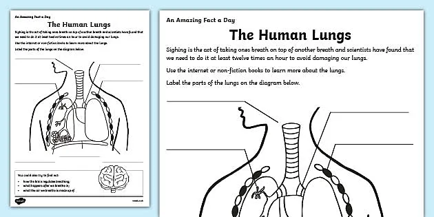
The Human Respiratory System You should be able to label a diagram showing: mouth, nose, trachea (windpipe), bronchi, bronchioles, alveoli (air sacs), pleural membranes, diaphragm, ribs and intercostal muscles. Use the mouse, or tap the screen, to label this diagram of the "complete " human respiratory system, combining the head and thorax (chest) regions. Respiratory system quizzes and labeled diagrams | Kenhub Take a look at the labeled diagram of the respiratory system above. As you can see, there are several structures to learn. Spend a few minutes reviewing the name and location of each one, then try testing your knowledge by filling in your own diagram of the respiratory system (unlabeled) using the PDF download below. Respiratory system unlabeled Complete the labeling of the diagram of the upper ... Complete the labeling of the diagram of the upper respiratory structures (sagittal section). \p iot go' P }''-'t ^r"\ hs;l Opening of pharyngotympanic tube IQ, Hyoid bone Thyroid gland \tP 2. ]1wo pairs of vocal folds are found in the larynx. Which pair are the true vocal cords (superior or inferior)? J ,.L rir. Nasopharynx Respiratory System - Comparative Anatomy Notes - 987 Words ... Complete the labeling of the diagram of the upper respiratory structures (sagittal section). Frontal sinus Cribriform plate of ethmoid bone Superior nasal chonchea middle inferior external nares Hard palate epiglottis Tongue Lingual tonsil tongue Hyoid bone Thyroid cartilage of larynx….
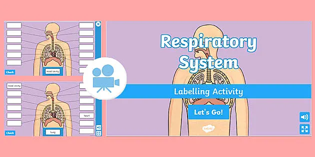
Respiratory System Labeling | Biology Game | Turtle Diary Respiratory System Labeling - Biology Game. Identify and label figures in Turtle Diary's fun online game, Respiratory System Labeling! Drag given words to the correct blanks to complete the labeling!
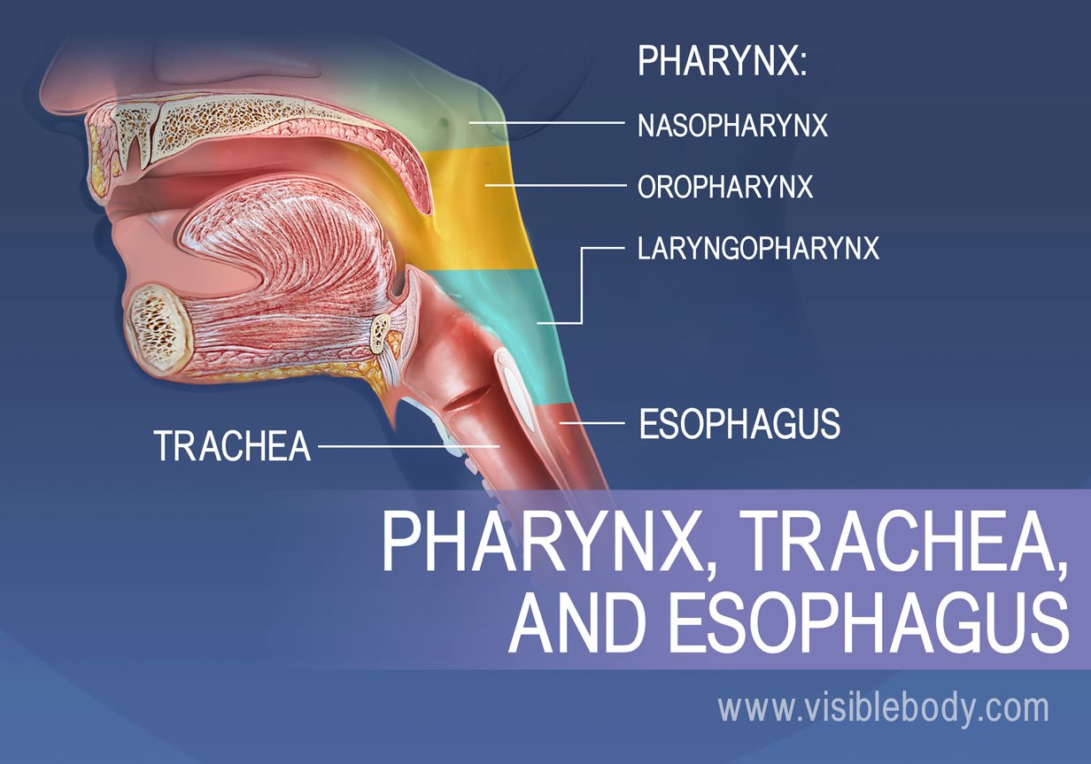
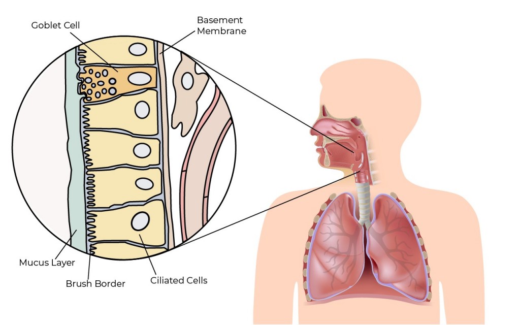
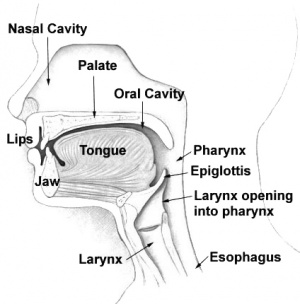
Upper Respiratory Tract: Anatomy, Functions, Diagram Upper Respiratory Tract Structural and Functional Anatomy Nose and Nasal Cavity The nostrils, the two round or oval holes below the external nose, are the primary entrance into the human respiratory system [5]. Lying just after the nostrils are the two nasal cavities, lined with mucous membrane, and tiny hair-like projections called cilia [6].
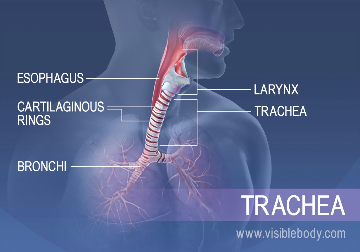
Larynx (Voice Box) Definition, Function, Anatomy, and Diagram The respiratory and digestive systems separate at the larynx, making it a vital organ in the function of both. Another primary function of the voice box is producing sounds and speech. Function in the respiratory system: Providing smooth passage of air from the nasal cavity to the lungs.
Diagram Of Larynx With Labeling Complete the labeling of the diagram of the upper respiratory structures (sagittal section). 2. Two pairs of vocal folds are found in the larynx. Which pair are the true vocal cords (superior or inferior)? 3. Name the specific cartilages in the larynx that correspond to the following descriptions.
Labeling Upper Respiratory Tract Diagram | Quizlet Label the structures of the upper respiratory tract. Learn with flashcards, games, and more — for free.
ZOO002L_EX014 respiratory.docx - ZOOLOGY LABORATORY ... ZOOLOGY LABORATORY EXERCISE 14: Upper and Lower Respiratory System Structures 1. Complete the labeling of the diagram of the upper respiratory structures (sagittal section).
Ex26_3rd ed.pdf - NAM LAB TIME/DATE_ Anatomy of the ... Complete the labeling of the diagram of the upper respiratory structures (sagittal section). Frontal sinus-.. Cribriform plate-of eth mold bone StLhPJtO1 --- cO& Sphenoidal sinus Opening of auditory tube Nasopharynx tv4erva-Y"--- Hard palate- -Iongue Hyoid bone- Thyroid cartilageof larynx Cricoid cartilage Thyroid gland yaLLfLcc - ___ .IOY1 U V 2.
PDF Label The Upper Respiratory System complete the labeling of the diagram of the upper respiratory structures sagittal section anatomy OF THE RESPIRATORY SYST'' The Respiratory System Diagram Functions Amp Organs May 2nd, 2018 - The Human Respiratory System Explained Including Anatomy Functions Organs With Diagrams Learning Aids And More '
Respiratory System Anatomy Labeling Diagram | Quizlet Also known as the voice box Trachea Also known as the windpipe; beginning of lower respiratory tract. Right bronchus Right branch off the trachea at the carina Left bronchus Left branch off the trachea at the carina Bronchiole Smaller airway dividing off the bronchi Diaphragm Main respiratory muscle Epiglottis
Amazing Detailed Respiratory System Diagram - Glaucoma ... The respiratory system is divided into an upper and lower respiratory tract. Students are to include all major components in their diagram design to include at minimum the 8 primary components of the system neatly labeling each aspect of the diagram in order to clearly communicate this idea. The nose and nasal cavity.
Solved 1. Complete the labeling of the diagram of the ... Question: 1. Complete the labeling of the diagram of the upper respiratory structures Frontal sinus Cribriform plate of ethmoid bone -Sphenoidal sinus -Opening of pharyngotympanic tube -Nasopharynx SI Hard palate Tongue the Hyoid bone Thyroid cartilage of larynx Cricoid cartilage Thyroid gland This problem has been solved! See the answer
Solved: Chapter E36 Problem 1E Solution | Human ... - Chegg Step 1 of 4 The respiratory tract is divided into two parts. They are upper respiratory tract and lower respiratory tract. Upper respiratory tract includes various passages and structures such as nose, nasal cavity, mouth, throat, and larynx (voice box). The entire respiratory tract is lined with a mucous membrane that secretes mucus.
Respiratory System - Science Quiz - Seterra Respiratory System - Science Quiz: Seterra is a free online quiz game that will teach you about science, biology, chemistry and the anatomy of the human body.
Upper Respiratory System (Labeling) Diagram | Quizlet Start studying Upper Respiratory System (Labeling). Learn vocabulary, terms, and more with flashcards, games, and other study tools.
Solved: Chapter E23 Problem 1E Solution | Laboratory ... Step 1 of 3 The respiratory tract is divided into two parts. They are upper respiratory tract and lower respiratory tract. Upper respiratory tract includes various passages and structures such as nose, nasal cavity, mouth, throat, and larynx (voice box). The entire respiratory tract is lined with a mucous membrane that secretes mucus.
Exercise 36 Review Sheet Anatomy of the Respiratory System ... Exercise 36 Review Sheet Anatomy of the Respiratory System Name Lab Time/DateUpper and Lower Respiratory System Structures 1 Complete the labeling of the diagram of the upper respiratory structures (sagittal section). 2 Two pairs of vocal folds are found in the larynx.
:background_color(FFFFFF):format(jpeg)/images/library/11020/respiratory_system_unlabeled.png)
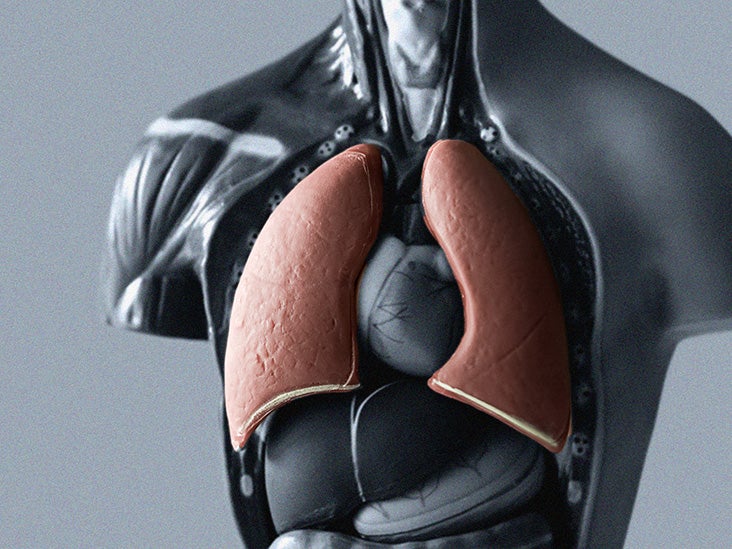


/human-respiratory-system-lungs-anatomy-953787016-b751ff559dc2489abdceb18b8fb77e8f.jpg)









0 Response to "40 complete the labeling of the diagram of the upper respiratory"
Post a Comment