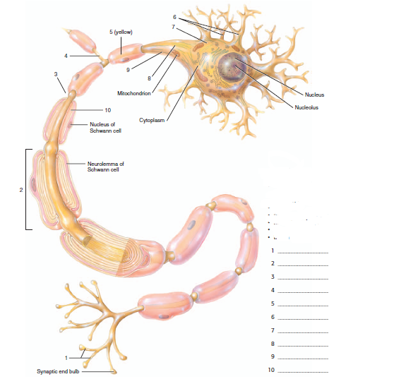
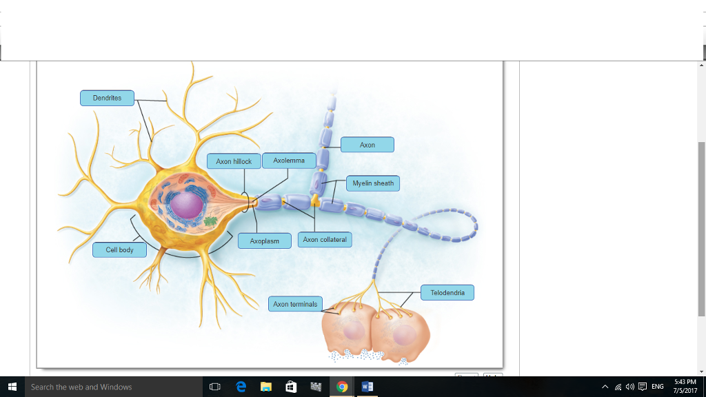
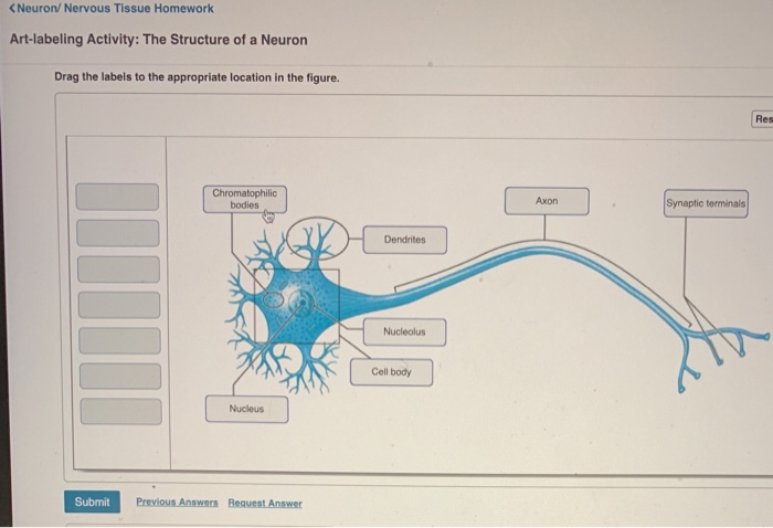
37 drag the labels onto the diagram to identify the parts of a myelinated pns neuron.
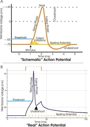
Nerves | Boundless Anatomy and Physiology A nerve is the primary structure of the peripheral nervous system (PNS) that encloses the axons of peripheral neurons. A nerve provides a structured pathway that supports neuron function. A nerve consists of many structures including axons, glycocalyx, endoneurial fluid, endoneurium, perineurium, and epineurium. Action potential - Definition, Steps, Phases - Kenhub An action potential is defined as a sudden, fast, transitory, and propagating change of the resting membrane potential. Only neurons and muscle cells are capable of generating an action potential; that property is called the excitability. This article will discuss the definition, steps and phases of the action potential.
PDF OpenALG OpenALG

Drag the labels onto the diagram to identify the parts of a myelinated pns neuron.
HW 7.pdf - HW 7 Due: 11:59pm on Friday, October 27, 2017 ... Part A Drag the labels onto the diagram to identify the parts of a myelinated PNS neuron. ANSWER: Correct Art-labeling Activity: Types of Neuroglia in the Central and Peripheral Nervous Systems Learning Goal: To learn the various types of neuroglia in the central and peripheral nervous systems. Labeled Neuron Diagram - Science Trends Dendrites: Dendrites are the receiving part of the neuron, the part that actually receives the electrical signal from other neurons. Dendrites look like long spindly extensions that branch out of the main body of the cell. In some kinds of neurons, dendrites themselves have further protrusions called dendritic spines that assist in electrical conductivity. Lab Exercise 12.pdf - 7/6/2021 Lab Exercise 12 Lab ... ANSWER: Correct Exercise 12 Pre-Lab Question 8 Part A From gross to microscopic, the parts of a muscle are _____. ANSWER: Correct Exercise 12 Review Sheet Art-labeling Activity 3 Identify the structures of the neuromuscular junction. Part A Drag the labels onto the diagram to identify the structures.
Drag the labels onto the diagram to identify the parts of a myelinated pns neuron.. NDSU Human Anat I- Exam 2 Flashcards - Quizlet Drag the resting membrane determinants to their appropriate locations in the figure. Answers may be used once, or not at all. Drag the appropriate labels to their targets. Note that pink labels should go in pink targets, blue labels should go in blue targets, and green labels should go in green targets. A&P2 Lab 13 HW, A&P2 Lab 12 HW, A&P2 Lab 11 HW ... - Quizlet Drag the labels onto the diagram to identify the parts of a myelinated PNS neuron. look at pic Drag the labels to identify the structural components of a typical synapse. Spinal Cord - Anatomy, Structure, Function, & Diagram Spinal Cord Anatomy. In adults, the spinal cord is usually 40cm long and 2cm wide. It forms a vital link between the brain and the body. The spinal cord is divided into five different parts. Several spinal nerves emerge out of each segment of the spinal cord. There are 8 pairs of cervical, 5 lumbar, 12 thoracics, 5 sacral and 1 coccygeal pair ... Stanford University UNK the , . of and in " a to was is ) ( for as on by he with 's that at from his it an were are which this also be has or : had first one their its new after but who not they have – ; her she ' two been other when there all % during into school time may years more most only over city some world would where later up such used many can state about national out known university united then made ...
Week 4 Chapter 13_.pdf - Week 4 Chapter 13 ... - Course Hero Part A Drag the labels to the appropriate location in the figure. ... In the peripheral nervous system, this myelin sheath is formed by the _____. Hint 1. ... Correct Part D - Structure of a Myelinated Neuron Label the diagram below by dragging and dropping the descriptions. Each label will be used only once. A Labelled Diagram Of Neuron with Detailed Explanations Learn More: Difference between Sensory and Motor Neuron. Diagram Of Neuron with Labels. Here is the description of human neuron along with the diagram of the neuron and their parts. The neuron is a specialized and individual cell, which is also known as the nerve cell. A group of neurons forms a nerve. Multipolar Neurons - Structure and Functions Multipolar neurons have three or more processes attached to the cell bodies. 1. 2. One process serves as the axon, which conducts electrochemical impulses (action potentials) between cells. 1. 2. The remaining processes are dendrites. Togather, the cell body and dendrites form the receptive zone of multipolar neurons. 1. Drag the labels onto the diagram to identify the ... Part A Drag the labels onto the diagram to identify the components of the somatic nervous system. ... About 90% of the parasympathetic fibers are located in which cranial nerve? the postganglionic neurons art in the PNS the neuron cell bodies are found in the CNS. the dote to the effectors. ... Drag the labels onto the diagram to identify the ...
Chapter 11 Homework Flashcards - Quizlet Drag the labels to identify the sequence of events that occurs at a synapse. Drag the labels onto the diagram to identify the various synapse structures. Arrange the parts in order, from left to right, of a successful direct depolarization path within one neuron. the receiving neuron is less likely to generate an action ... Label the events of presynaptic inhibition and facilitation. Part A Drag the labels onto the diagram to identify the events of presynaptic inhibition and facilitation. ANSWER: ependymal cells Schwann cells satellite cells astrocytes saltatory hyperpolarization graded continuous acetylcholine dopamine norepinephrine gamma aminobutyric acid (GABA) Solved Art-labeling Activity: Parts of a Myelinated | Chegg.com These are the different parts of neurons. It is an electrically excitable cell. It carries inf …. View the full answer. Transcribed image text: Art-labeling Activity: Parts of a Myelinated Peripheral Nervous System (PNS) Neuron Drag the labels onto the diagram to identify the parts of a myelinated PNS neuron. Nodes. Central nervous system - Wikipedia The central nervous system (CNS) is the part of the nervous system consisting primarily of the brain and spinal cord.The CNS is so named because the brain integrates the received information and coordinates and influences the activity of all parts of the bodies of bilaterally symmetric and triploblastic animals—that is, all multicellular animals except sponges and diploblasts.
m8ta fun
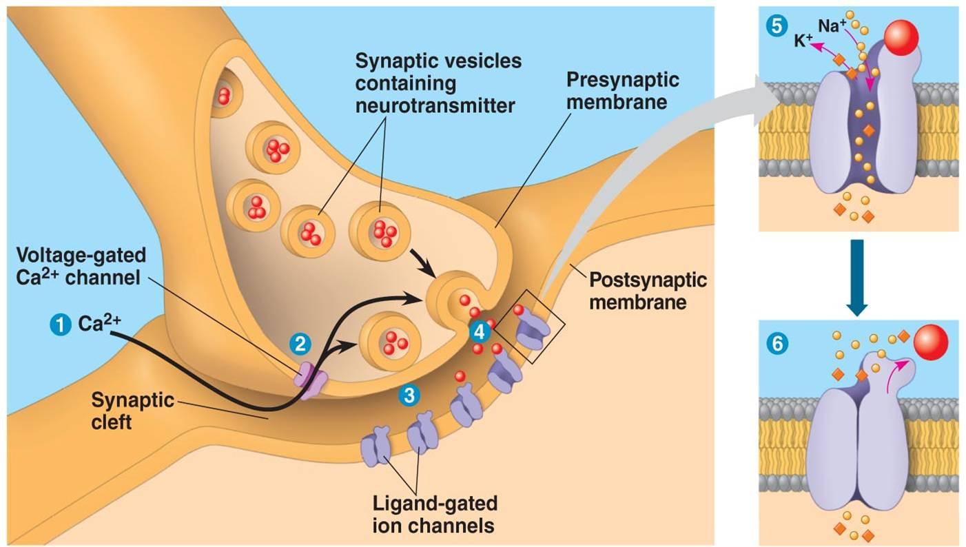
PDF The Autonomic Nervous System and Visceral Sensory Neurons municates the message from one neuron, called the presyn-aptic neuron, to another neuron, the postsynaptic neuron. A ganglion (plural: ganglia) is a cluster of neuronal cell bodies in the PNS. (See Chapter 12 for review of these terms.) Comparison of the Autonomic and Somatic Motor Systems Other discussions of motor innervation focus largely on the

A&P2 Lab 1 HW Flashcards | Quizlet
PDF The world's learning company | Pearson The world's learning company | Pearson

12.2 Nervous Tissue – Anatomy & Physiology
Structure of the Autonomic Nervous System | Boundless ... The enteric nervous system is also sometimes considered part of the autonomic nervous system, and sometimes considered an independent system. The ANS is unique in that it requires a sequential two- neuron efferent pathway; the preganglionic neuron must first creat a synapse to a postganglionic neuron before innervating the target organ.

Lecture 14 (Chapter 13) Spinal Cord. - ppt download
A&P2 Lab 1 HW Flashcards - Quizlet Drag the labels onto the diagram to identify the parts of a myelinated PNS neuron. look at pic Drag the labels to identify the structural components of a typical synapse.

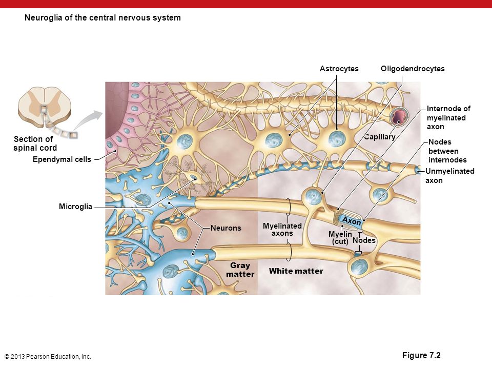
Neuroglia | Boundless Anatomy and Physiology
Location, Structure, and Functions of Sensory ... - Bodytomy The CNS is made of the brain and spinal cord. The PNS consists of nerves, and it connects the CNS to the organs of the periphery. Neurons are basic functional units of the nervous system. Their function is to conduct nerve impulses. Depending on the type of impulse they conduct, neurons can be classified into sensory neurons, motor neurons, or ...

HW 7.pdf - HW 7 Due: 11:59pm on Friday, October 27, 2017 To ...
drag the labels into the diagram to identify the ... Part A Drag the labels onto the diagram to identify the parts of the corticospinal pathway. Reset Help rary Motor Corte Cerebral peduncle Medulla oblongata Anterior corticospinal tract To Lateral corticospinal tract skeletal muscles Midbrain Το Spinal cord skeletal muscles Motor nuclei of cranial nerves Corticobulbar tract Crossover of ...
The Nervous System
A&P2 Lab and Powerpoints Flashcards - Quizlet Drag the labels onto the diagram to identify the parts of a myelinated PNS neuron. look at pic Drag the labels to identify the structural components of a typical synapse.

Brain HW Flashcards | Quizlet
Solved Drag the labels onto the diagram to identify the ... Drag the labels onto the diagram to identify the parts of a myelinatwed PNS neuron; Question: Drag the labels onto the diagram to identify the parts of a myelinatwed PNS neuron. This problem has been solved! See the answer See the answer See the answer done loading. Show transcribed image text
Ch 11 Nervous system Flashcards | Chegg.com
Lab Exercise 12.pdf - 7/6/2021 Lab Exercise 12 Lab ... ANSWER: Correct Exercise 12 Pre-Lab Question 8 Part A From gross to microscopic, the parts of a muscle are _____. ANSWER: Correct Exercise 12 Review Sheet Art-labeling Activity 3 Identify the structures of the neuromuscular junction. Part A Drag the labels onto the diagram to identify the structures.

Comparative Anatomy of Glial Cells in Mammals - ScienceDirect
Labeled Neuron Diagram - Science Trends Dendrites: Dendrites are the receiving part of the neuron, the part that actually receives the electrical signal from other neurons. Dendrites look like long spindly extensions that branch out of the main body of the cell. In some kinds of neurons, dendrites themselves have further protrusions called dendritic spines that assist in electrical conductivity.
BTEC International Level 3 Qualifications in Applied Science ...
HW 7.pdf - HW 7 Due: 11:59pm on Friday, October 27, 2017 ... Part A Drag the labels onto the diagram to identify the parts of a myelinated PNS neuron. ANSWER: Correct Art-labeling Activity: Types of Neuroglia in the Central and Peripheral Nervous Systems Learning Goal: To learn the various types of neuroglia in the central and peripheral nervous systems.
Anatomy Exam 2 Flashcards - Easy Notecards
Neuron Labeling Worksheets & Teaching Resources | TpT
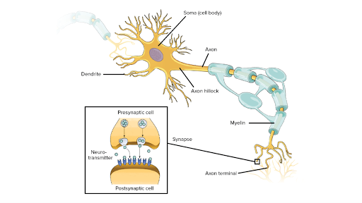
Overview of neuron structure and function (article) | Khan ...
Neuron Labeling Worksheets & Teaching Resources | TpT
Solved Dendrites Axon Axon hillock Axolemma Myelin sheath ...

4.1 The Neuron Is the Building Block of the Nervous System ...

Comparative Anatomy of Glial Cells in Mammals - ScienceDirect
Synaptic Transmission between Neurons | Neupsy Key

NDSU Human Anat I- Exam 2 Flashcards | Quizlet

NDSU Human Anat I- Exam 2 Flashcards | Quizlet
Nervous System
Threshold potential - Wikipedia

A&P2 Lab 1 HW Flashcards | Quizlet

12.2 Nervous Tissue – Anatomy & Physiology

Healthmedicinet Com II 2014 Sep | PDF | Ebola Virus Disease ...
https://www.frontiersin.org/articles/10.3389/fnins.2013.00093 ...

Biol 2022 Nervous System Flashcards | Quizlet

Ch 12 - Nervous System EXAM *** McGraw Flashcards | Quizlet
Solved Exercise 14 Review Sheet: Nervous Tissue Drag the ...
11 Introduction to the Nervous System and Nervous Tissue
PowerPoint ® Lecture Slides prepared by Betsy C. Brantley ...
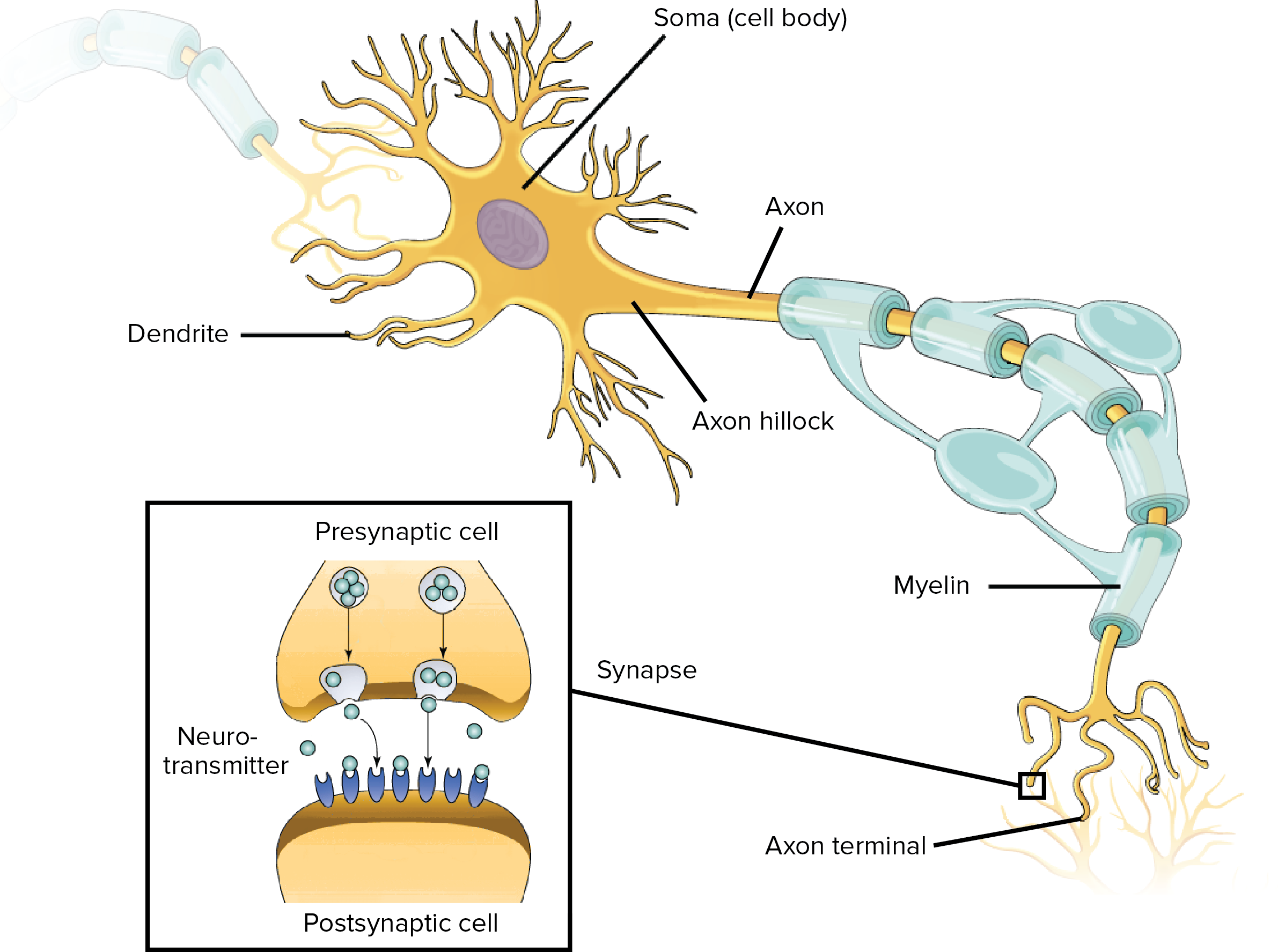
Overview of neuron structure and function (article) | Khan ...
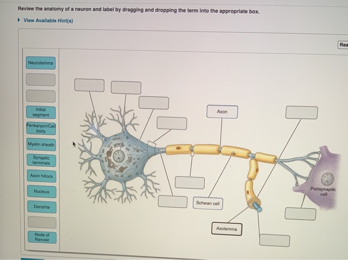
Solved Review the anatomy of a neuron and label by dragging ...

Which nerve arises from the sacral plexus o Sciatic nerve ...

12.2 Nervous Tissue – Anatomy & Physiology

Parts of a Myelinated Peripheral Nervous System (PNS) Neuron ...
0 Response to "37 drag the labels onto the diagram to identify the parts of a myelinated pns neuron."
Post a Comment