38 human cheek cell diagram
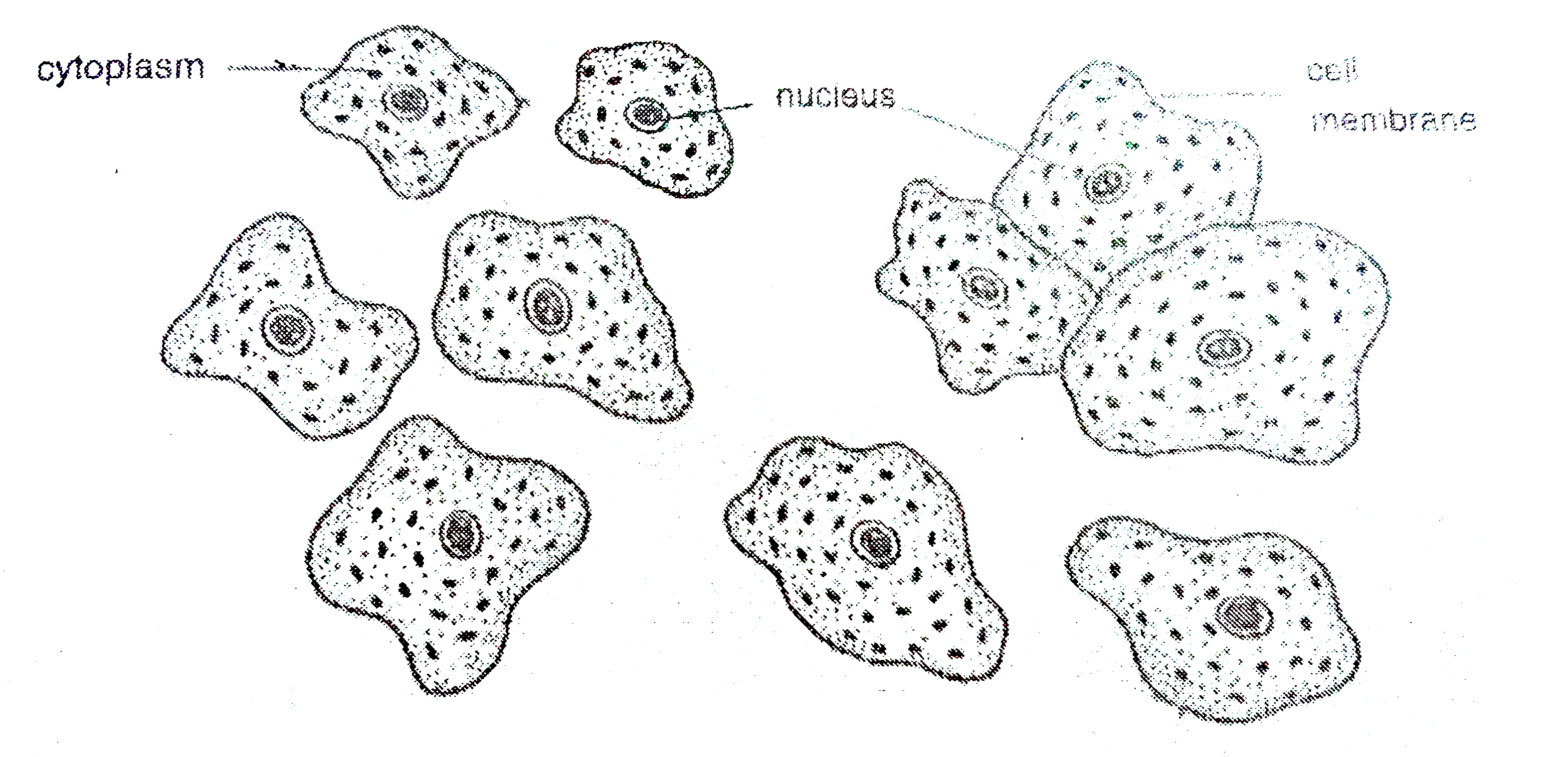
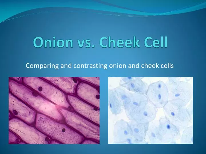
Difference Between Onion Cell and Human Cheek Cell ... The human cheek cell is a type of animal cell and it can be obtained by scraping the cheek from a toothpick. It is made up of simple squamous epithelium. Since human cheek cells are animal cells they do not have a cell wall. Hence, the outside barrier of the human cheek cell is the cell membrane, which serves as the semi-permeable barrier. Onion and Cheek Cells (Theory) : Class 9 : Biology ... Human Cheek Cell As in all animal cells, the cells of the human cheek do not possess a cell wall. A cell membrane that is semi-permeable surrounds the cytoplasm. Unlike plant cells, the cytoplasm in an animal cell is denser, granular and occupies a larger space. The vacuole in an an animal cell is smaller in size, or absent.
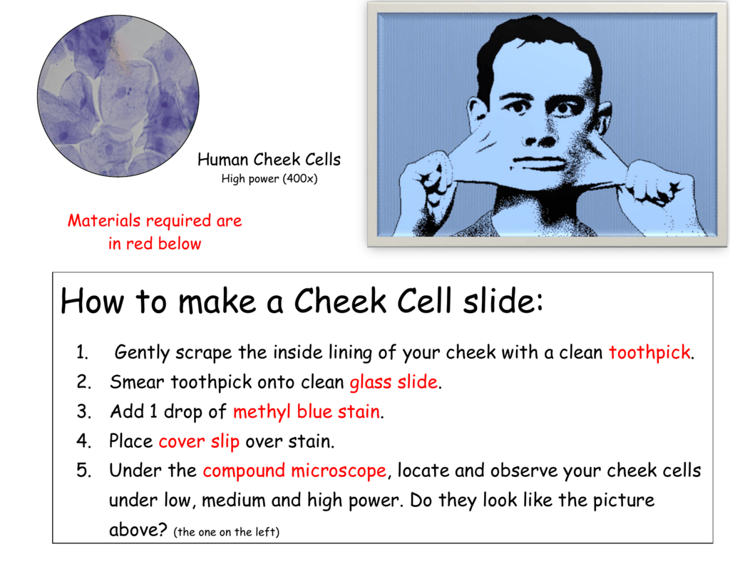
Human Cheek - Experiments on Microscopes 4 Schools Human cheek cells Materials Glass microscope slides Plastic cover slips Paper towels or tissue Methylene Blue solution (0.5% to 1% (mix approximately 1 part stock solution with 4 parts of water)) Plastic pipette or dropper Sterile, individually packed cotton swabs See information on suppliers here. Methods

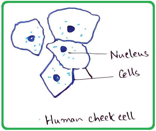
Human cheek cell diagram
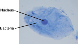
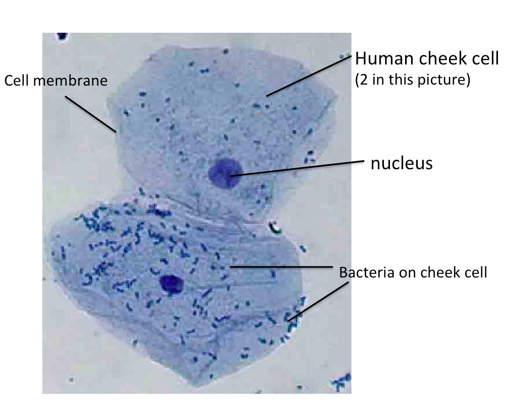
Deoxyribonucleic Acid (DNA) Fact Sheet - Genome.gov 24.08.2020 · When a cell prepares to divide, the DNA helix splits down the middle and becomes two single strands. These single strands serve as templates for building two new, double-stranded DNA molecules - each a replica of the original DNA molecule. In this process, an A base is added wherever there is a T, a C where there is a G, and so on until all of the bases once again have … To prepare stained temporary mounts of human cheek cell ... 1. Cheeks should be scrapped gently to prevent injury. 2. Spread the material on the slide so that it forms a thin uniform layer. 3. Avoid over staining (or under staining) of the material. 4. While mounting the cover slip, avoid entry of air bubbles. Go to List of Experiments Previous Post Next Post Cell Structure - Northern Kentucky University This human cheek cell is a good example of a typical animal cell. It has a prominent nucleus and a flexible cell membrane which gives the cell its irregular, soft-looking shape. Like most eukaryotic cells, this cell is very large compared to prokaryotic cells.
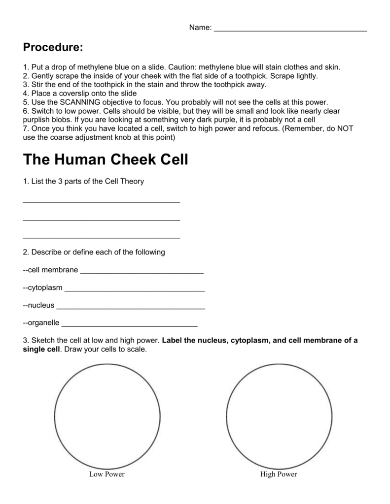
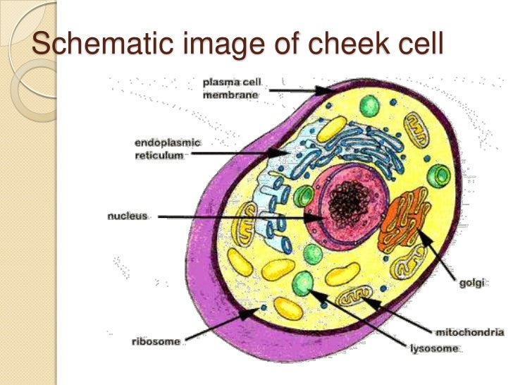
Human cheek cell diagram. Submandibular Lymph Nodes Anatomy, Diagram & Function ... 20.01.2018 · The submandibular lymph nodes sit between the submandibular salivary glands, which are underneath the tongue, and the mandible, or lower jawbone. Occasionally one or more of the lymph nodes may be ... How to draw Human Cheek Cell/2019 - YouTube #How to draw Human Cheek Cell Cell Nucleus - function, structure, and under a microscope ... [In this figure] The cell nucleus diagram and its structure. ... Are there human diseases associated with nucleus dysfunction. The nucleus is the hallmark of eukaryotic cells; therefore, the structural proteins in the nuclear envelope are important for maintaining the nuclear function. The mutations in the nuclear envelope such as lamins cause a number of human diseases, … PDF Biological drawing of cheek cell light purple liquid in the cell. The sample was then stained with methylene blue so the cells could be viewed at 40x 100x and 400x. Draw A Neat Diagram Of Human Cheek Cell And Label Its Three Parts Brainly In Chapter 1 page 7 histologyolm. Human cheek cell labeled diagram. Cheek cells are eukaryotic cells cells that contain a nucleus and other
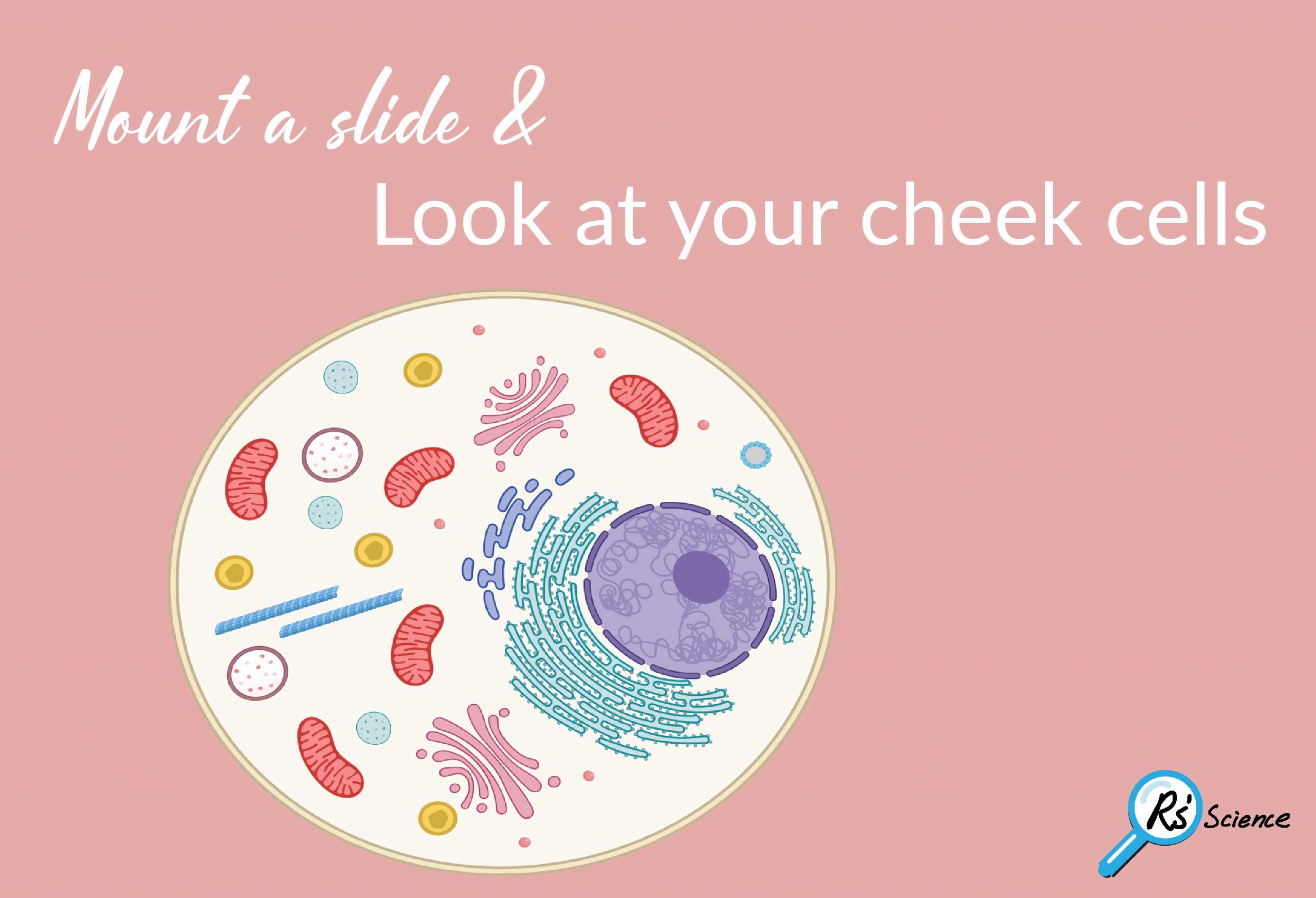
Cheek Cells Under a Microscope - Requirements/Preparation ... Cheek cells are eukaryotic cells (cells that contain a nucleus and other organelles within enclosed in a membrane) that are easily shed from the mouth lining. It's therefore easy to obtain them for observation. Some of the main parts of a cell include: 1. Cell membrane (outer boundary of the cell) 2. Cytoplasm (the fluid within the cell) 3. Comparing sizes - Cell structure - AQA - GCSE Combined ... The diagram shows the size of three organisms, different cells and other structures. Sizes can be compared using a straightforward calculation. For instance, the length of the leaf cell above is ... Human Cheek Epithelial Cells - Florida State University Human Cheek Epithelial Cells The tissue that lines the inside of the mouth is known as the basal mucosa and is composed of squamous epithelial cells. These structures, commonly thought of as cheek cells, divide approximately every 24 hours and are constantly shed from the body. Human Cell Diagram, Parts, Pictures, Structure and ... Diagram of the human cell illustrating the different parts of the cell. Cell Membrane The cell membrane is the outer coating of the cell and contains the cytoplasm, substances within it and the organelle. It is a double-layered membrane composed of proteins and lipids.
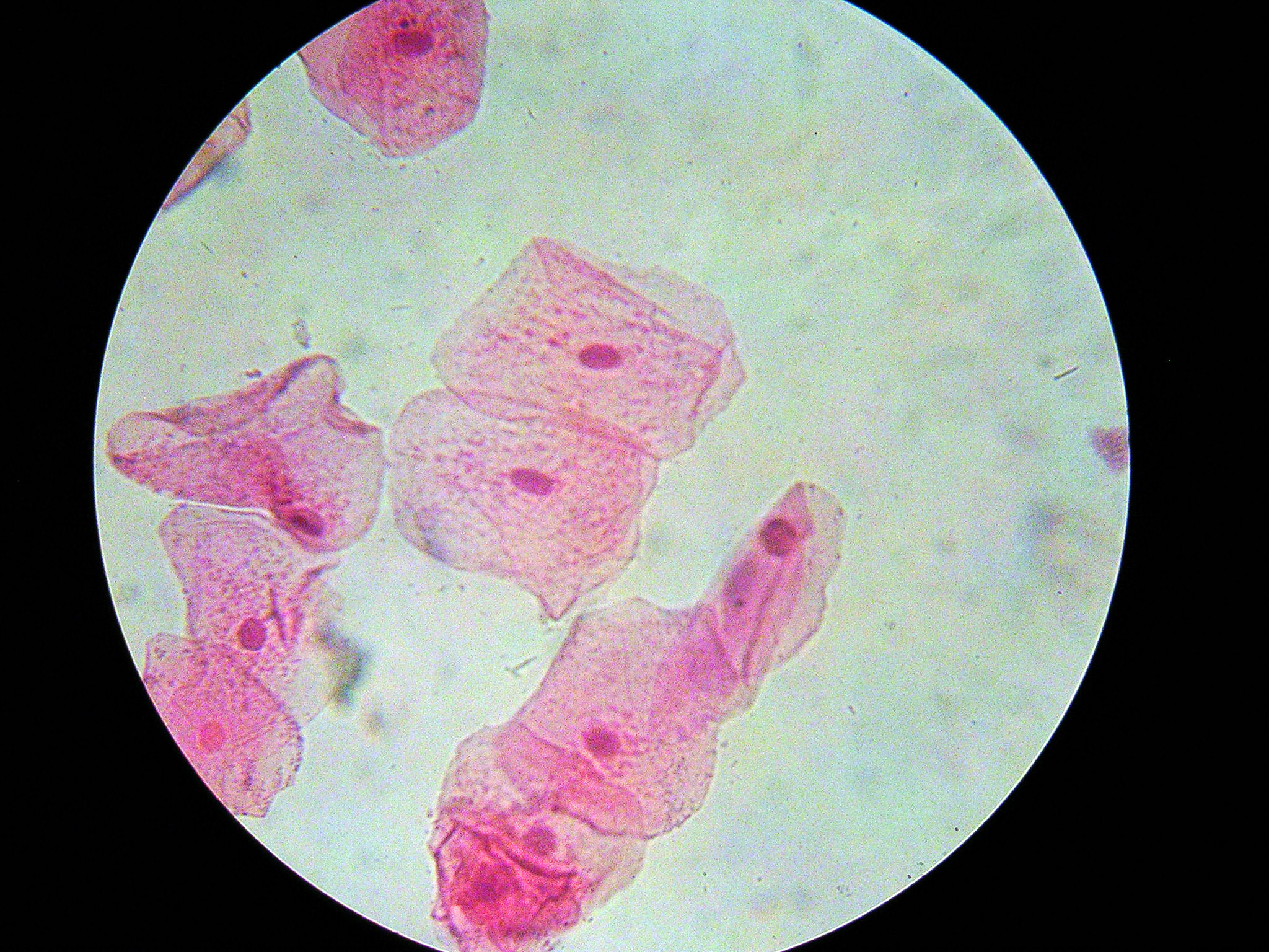
PDF Lab 3: Cells: Structure and Function Draw a well-labeled diagram of the cheek cells on the paper provided. Be sure to provide the magnification, and estimate the size of the cells, and the size of the nucleus. Plant Cells: ElodeaCells Now you will examine a typical plant cell, from the leaf of the aquatic plant Elodea. 1. Gently remove one leaf from near the tip of an Elodeastalk. 2. Human Cheek Cells Under the Microscope | Haematoxylin ... Human cheek cells are observed under microscope- 1. Cells are polygonal or flat in shape and structure - 2. They have irregular cellular thin boundaries which contains jelly like cytoplasm and the cytoplasm are granular. 3. This cell do not have plastids, vacuoles or cell wall. 4. They are generally made up of squamous epithelium cells. PDF Human Cell Diagram, Parts, Pictures, Structure and Functions Diagram of the human cell illustrating the different parts of the cell. Cell Membrane The cell membraneis the outer coating of the cell and contains the cytoplasm, substances within it and the organelle. It is a double-layered membrane composed of proteins and lipids. PDF Cell Structure - University of Massachusetts Boston Animal cells Human cheek cells (4.0 x 10²x) Photograph: Y. Vaillancourt, UMass Boston, 2006 Guinea pig pancreas (6.0 x 10⁴x) Photograph : Dr. George Palade, Rockefeller Institute, NY Cell Ultrastructure, Jensen and Park, 1967 Guinea pig pancreas (3.0 x 10⁴x) PPhotograph : hotograph : DDr.
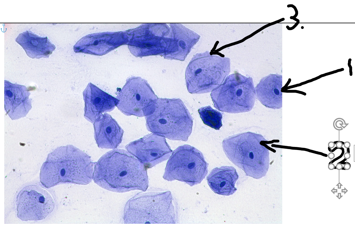
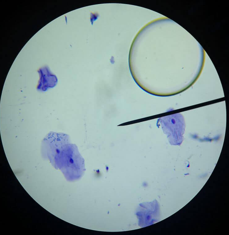
Viewing Human Cheek Cells - The Biology Corner Viewing Human Cheek Cells In this lab, we took a sample of our own cells by scraping the inside of the cheek with a toothpick. The sample was then stained with methylene blue, so the cells could be viewed at 40x, 100x, and 400x. The following images simulate what would be seen at each magnification. Cheek cells 40x. Cheek Cells, 100x
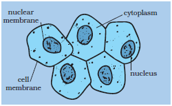
Science 8 Final Exam Review 2019 | Science Quiz - Quizizz A student was studying a prepared slide of human cheek cells under a compound light microscope. The diagram represents what the student observed on the slide at a magnification of 100×. Identify the shaded structure shown in each cell in the diagram.
What are the parts of cheek cell? - Rehabilitationrobotics.net What is a human cheek cell? The tissue that lines the inside of the mouth is known as the basal mucosa and is composed of squamous epithelial cells. These structures, commonly thought of as cheek cells, divide approximately every 24 hours and are constantly shed from the body.
Which of the following is the diagram of human cheek cell? Which of the following is the diagram of human cheek cell? A B C D Medium Solution Verified by Toppr Correct option is A As in all animal cells, the cells of the human cheek do not possess a cell wall. A cell membrane that is semi-permeable surrounds the cytoplasm. The vacuole in an animal cell is smaller in size, or absent.
unit II IX b - NCERT To prepare a temporary mount of human cheek epithelial cells, and to study its characteristics. Like plants, the body of all animals including humans is composed of cells. Unlike plant cells, animal cells do not have cell wall. The outermost covering of an animal cell is a cell membrane. The cytoplasm, nucleus and other cell organelles are enclosed in it. Epithelial tissue is the …
Structure of human cheek cell? - Answers The cheeks cell in the human body is to do everything shown on the out side of the cheek.For example if you are sticking in your cheek or jaw in, the cell in the cheek is working and doing the ...
Cheek Cells Lesson Plans & Worksheets Reviewed by Teachers Human Cheek Cell. For Students 8th - 10th. Get up close and personal with human cells with this lab worksheet. Learners use a microscope to examine their own cheek cells, drawing diagrams of the cells and identifying the parts when they have focused in on a visible specimen.... Get Free Access See Review.
PPT Cheek Cell - YCSD Our cheek cells are clear. Iodine is a brown color. It is also a stain. I will turn our cheek cells a brown color so that we will see them. 6. The light microscope used in the lab is NOT powerful enough to view other organelles in the cheek cell. What parts of the cell were visible. Cell membrane Cytoplasm You might have seen nucleus 6.
Human eye - Wikipedia The human eye is a sense organ, part of the sensory nervous system, that reacts to visible light and allows us to use visual information for various purposes including seeing things, keeping our balance, and maintaining circadian rhythm.. The eye can be considered as a living optical device.It is approximately spherical in shape, with its outer layers, such as the outermost, white …
courseworkhero.co.ukCoursework Hero - We provide solutions to students We provide solutions to students. Please Use Our Service If You’re: Wishing for a unique insight into a subject matter for your subsequent individual research;
Human tooth - Wikipedia Diagram of a human molar showing its major constituents. Details; Identifiers; Latin: dentes: TA98: A05.1.03.001: TA2: 2818: FMA: 75150: Anatomical terminology [edit on Wikidata] The human teeth function to mechanically break down items of food by cutting and crushing them in preparation for swallowing and digesting. Humans have four types of teeth: incisors, canines, …
What Is the Function of the Cheek Cell? - Reference.com A human cheek cell is thin, flat and irregularly shaped and has a large nucleus that contains the DNA. Its plasma membrane helps the cell to maintain suitable temperature while giving it its shape. Since it is selectively permeable, it only allows certain molecules into and out of the cell.
Diagram of human cheek cell and onion cell - Brainly.in Human cheek cells - A cell layer that is semi-porous encompasses the cytoplasm. Human cheek cells are adjusted. Human cheek cells don't have a cell divider or a huge vacuole Both onion and human cheek cells are epithelial cells. Onion Cells -Onion cells are block like in shape. An onion is a multicellular (comprising of numerous cells) plant ...
bio.libretexts.org › Bookshelves › Human_Biology23.3: Embryonic Stage - Biology LibreTexts The epidermis, glands of the skin, some cranial bones, pituitary and adrenal medulla, the nervous system, the mouth between cheek and gums, the anus: Mesoderm: Connective tissues, bone, cartilage, blood endothelium of blood vessels, muscles, synovial membranes, serous membranes, kidneys, the lining of gonads: Endoderm
patapum.to.itFlour Mill Rye [4MH368] Search: Rye Flour Mill. What is Rye Flour Mill. Every flour has its own unique properties. Sourdough Rye using your flour and some crushed organic caraway seeds has lifted my Sourdough Rye to a new level!!
SCIENCE (5 2) - CISCE human welfare. 5. To understand the capacities and limitations of all the biological and economic activities so as to be able to use them for a better quality of life. 6. To acquire the ability to observe, experiment, hypothesize, infer, handle equipment accurately and make correct recordings. CLASS IX There will be one paper of two hours duration of 80 marks and Internal …
› lakhmir-singh-science-class-8Lakhmir Singh Science Class 8 Solutions Chapter 8 Cell ... Jun 11, 2019 · Draw the general diagram of an animal cell and label it. Answer: Question 68 B. Draw the general diagram of a plant cell and label it. Answer: Question 68 C. Explain why, chloroplasts are found only in plant cells. Answer: Chloroplasts are the green coloured plastids present in the cytoplasm of plant cells.
PHOTOSYNTHESIS - Estrella Mountain Community College Diagram of a typical plant, showing the inputs and outputs of the photosynthetic process. Image from Purves et al., Life: The Science of Biology, 4th Edition, by Sinauer Associates ( ) and WH Freeman ( ), used with permission. Leaves and Leaf Structure | Back to Top. Plants are the only photosynthetic organisms to have leaves (and …
NCERT Class 9 Science Lab Manual - Slide of ... - CBSE Tuts human cheek cells and to record observations and draw their labelled diagrams. Theory Plant cell to be studied in lab: Onion peel The cells are very clearly visible as compartments with prominent nucleus in it. The cell walls are very distinctly seen under the microscope. The big vacuoles are also seen in each cell.
how to draw human cheek cell diagram | how to draw cheek ... Title: how to draw human cheek cell | how to draw human cheek cell easily | how to draw human cheek cellHello Friends in this video I tell you about how to d...
UNIT 3 - NCERT You have earlier observed cells in an onion peel and/or human cheek cells under the microscope. Let us recollect their structure. The onion cell which is a typical plant cell, has a distinct cell wall as its outer boundary and just within it is the cell membrane. The cells of the human cheek have an outer membrane as the delimiting structure of the cell. Inside each cell …
Practical Reports./ Onion Cells/ Human Cheek Cells/ Fungal ... 2. Cell Structure Practical Exercises: ( 3 Parts in one report). ( 2-3 pages) a. Onion Cells b. Human Cheek Cells c. Fungal Cells. Biology 2. Cell Structure Practical Exercises. Introduction. In this practicalexercise you will look at three different categories of cell: Fungal, Animal and Plant.
Human Cheek Cell Diagram Labeled - Diagram Sketch Human Cheek Cell Diagram Labeled angelo on September 18, 2021 Label The Plant Cell Worksheets Sb11867 Sparklebox Cells Worksheet Plant Cell Plant Cells Worksheet Art Animal Cells Do Not Have Cell Walls They Can Change Size And Shape More Easily Than Plant Cells Animal Cell Plant And Animal Cells Animal Cell Project
Which of the following is the diagram of human cheek cell? Which of the following is the diagram of human cheek cell? A B C D Answer Correct option is A As in all animal cells, the cells of the human cheek do not possess a cell wall. A cell membrane that is semi-permeable surrounds the cytoplasm. The vacuole in an animal cell is smaller in size, or absent.
Cell Division 1 QP.pdf - Q1.Figure 1 shows a human cheek ... Page 14 (a) The diagram shows a yeast cell. Parts A and B are found in human cells and in yeast cells. On the diagram, label parts A and B. (2) (b) Many types of cell can divide to form new cells. Some cells in human skin can divide to make new skin cells.
Cell Structure - Northern Kentucky University This human cheek cell is a good example of a typical animal cell. It has a prominent nucleus and a flexible cell membrane which gives the cell its irregular, soft-looking shape. Like most eukaryotic cells, this cell is very large compared to prokaryotic cells.
To prepare stained temporary mounts of human cheek cell ... 1. Cheeks should be scrapped gently to prevent injury. 2. Spread the material on the slide so that it forms a thin uniform layer. 3. Avoid over staining (or under staining) of the material. 4. While mounting the cover slip, avoid entry of air bubbles. Go to List of Experiments Previous Post Next Post
Deoxyribonucleic Acid (DNA) Fact Sheet - Genome.gov 24.08.2020 · When a cell prepares to divide, the DNA helix splits down the middle and becomes two single strands. These single strands serve as templates for building two new, double-stranded DNA molecules - each a replica of the original DNA molecule. In this process, an A base is added wherever there is a T, a C where there is a G, and so on until all of the bases once again have …













0 Response to "38 human cheek cell diagram"
Post a Comment