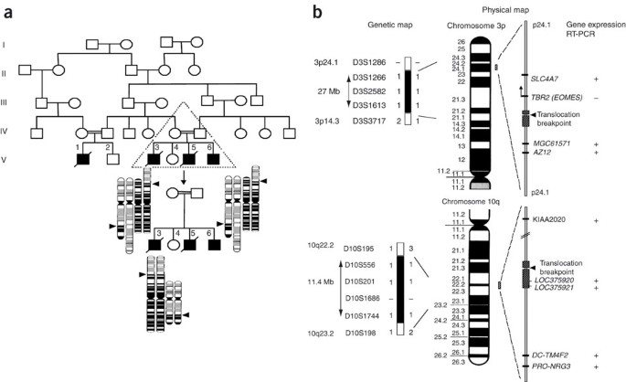
36 figure 11-4 is a diagram of the frontal section of the heart
DOC Dorchester School District Two / Homepage Figure 11-4 is a diagram of the frontal section of the heart. Follow the instructions below to complete this exercise. (a) Correctly identify each of the heart valves (numbers 6-9 on the figure) by inserting the appropriate terms in the blanks left of the figure. (b) Use the numbers from the figure to identify the structures described below. Ch. 14 Introduction - Anatomy and Physiology - OpenStax Ch. 14 Introduction - Anatomy and Physiology | OpenStax. Figure 14.1 Too Hot to Touch When high temperature is sensed in the skin, a reflexive withdrawal is initiated by the muscles of the arm. Sensory neurons are activated by a stimulus, which is sent to the central nervous system, and a motor response is sent out to the skeletal muscles that ...
Diagram of Human Heart and Blood Circulation in It | New ... A heart diagram labeled will provide plenty of information about the structure of your heart, including the wall of your heart. The wall of the heart has three different layers, such as the Myocardium, the Epicardium, and the Endocardium. Here's more about these three layers. Epicardium

Figure 11-4 is a diagram of the frontal section of the heart
PDF Home - Buckeye Valley Created Date: 1/23/2014 12:31:42 PM openstax.org › books › anatomy-and-physiology1.6 Anatomical Terminology - Anatomy and Physiology - OpenStax Figure 1.14 Planes of the Body The three planes most commonly used in anatomical and medical imaging are the sagittal, frontal (or coronal), and transverse plane. Body Cavities and Serous Membranes The body maintains its internal organization by means of membranes, sheaths, and other structures that separate compartments. NUMC 101 Module 2 Cardiovascular System Heart Lab Exercise ... Figure 11-4 is a diagram of the frontal section of the heart. Follow the instructions below to complete this exercise. First, draw arrows to indicate the direction of blood flow through the heart. Draw the pathway of the oxygen-rich blood with red arrows, and trace the pathway of the oxygen-poor blood with blue arrows.
Figure 11-4 is a diagram of the frontal section of the heart. PDF Anatomy of the Heart Anatomy of the Heart Objectives: 1. To describe the location of the heart. 2. To name and locate the major anatomical areas and structures of the heart when provided with an appropriate model or diagram, or with a dissected sheep heart, and to explain the function of each. 3. To trace the pathway of blood through the heart. 4. › 31489704 › the_handbook_ofthe_handbook_of_highway_engineering.pdf - Academia.edu Academia.edu is a platform for academics to share research papers. PDF Cardiovascular System Components of the ... - soinc.org The average heart beats 100,000 times per day pumping about 2,000 gallons (7,571 liters) of blood. • The average human heart beating at 72 BPM (beats per minute), will beat approximately 2.5 billion times during a lifetime of 66 years. • The heart is usually situated in the middle of the thorax with the largest part of the heart slightly Heart Anatomy: Labeled Diagram, Structures, Blood Flow ... Image: Cartoon diagram of the heart we will use to review the main cardiac structures and anatomy. Chambers of the Heart Let's begin with the chambers of the heart. There are 4 chambers, labeled 1-4 on the diagram below. To help simplify things, we can convert the heart into a square.
Figure 38.3 Frontal section of human heart Diagram | Quizlet Start studying Figure 38.3 Frontal section of human heart. Learn vocabulary, terms, and more with flashcards, games, and other study tools. (Get Answer) - Look at the model. The arrows indicate the ... 209 Chapter 11 The Cardiovascular System 6. Figure 11-4 is a diagram of the frontal section of the heart instructions below to complete this exercise Follow the First, draw arrows to indicate the direction Draw the pathway of the b the of blood flow... PDF M11 MARI5326 12 SE C11.indd Page 356 24/09/16 9 ... - Pearson of the blood vessels leaving and entering the heart. (Figure 11.3 shows two views of the heart—an exter-nal anterior view and a frontal section. As the ana-tomical areas of the heart are described in the next section, keep referring to Figure 11.3 to locate each of the heart structures or regions.) Chambers and Associated Great Vessels Heart Anatomy Frontal Section Diagram | Quizlet Brachiocephalic Artery The first major branch off of the aorta and the major artery to the forelimbs and head. Superior Vena Cava receives blood from the head and arms and chest and empties into the right atrium of the heart Ascending Aorta the ascending part of the aorta as it emerges from the left ventricle Pulmonary Semilunar Valve
PDF The Urinary System - Pearson A frontal section of the kidney reveals the three distinct regions of this organ: the outermost renal cortex, the middle renal medulla, and the inner renal pelvis (Figure 24.3a). Together, the renal cort ex and the renal medulla make up the urine-forming portion of the kidney. The renal pelvis and associated structures drain urine that › 11207373 › Ashrae_1999_HVACAshrae 1999 HVAC Applications Handbook - Academia.edu Academia.edu is a platform for academics to share research papers. Solved 6. Figure 11-4 is a diagram of the interior frontal ... Question: 6. Figure 11-4 is a diagram of the interior frontal section of the heart. (A) Draw arrows to indicate the direction of blood flow through the heart and great vessels. (B) Color the heart chambers and the vessels transporting O2-poor blood blue and chambers and vessels transporting O2-rich blood red. 10 Chapter 11 The Cardiovascular System 6 Figure 114 is a ... Chapter 11 The Cardiovascular System 6. Figure 11—4 is a diagram of the frontal section of the heart. instructions below to complete this exercise.
Frontal Section of the Heart Diagram Diagram | Quizlet
PDF The Cardiovascular System - Pearson of the blood vessels leaving and entering the heart. (Figure 11.3 shows two views of the heart—an exter-nal anterior view and a frontal section. As the ana-tomical areas of the heart are described in the next section, keep referring to Figure 11.3 to locate each of the heart structures or regions.) Chambers and Associated Great Vessels
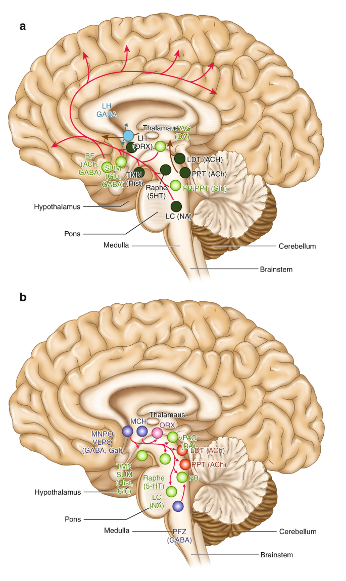
Neurobiology of Insomnia | SpringerLink
Cardiovascular System - Heart - Building a Medical ... Figure 12.6 image description: This image shows the anterior view of the frontal section of the heart with the major parts labeled. Labels read (from top of diagram, clockwise) arch of aorta, Bachman's bundle, atrioventricular bundle (bundle of His), left ventricle, right and left bundle branches, Purkinje fibers, right ventricle, right ...
Handbook of Operational and Aerospace Physiology
pharmmedexpert.de › thigh-measurements-for-size-0Thigh measurements for size 0 - pharmmedexpert.de Care. Put 'em together and the example bra size is 34B. The thigh measurement is used for the swimwear. From the frontal and lateral measurements obtained, the waist, lower hip, and thigh areas were calculated, which were considered to be ellipse-shaped [15]. Boohoo. Measure your chest and hips at the largest size area.
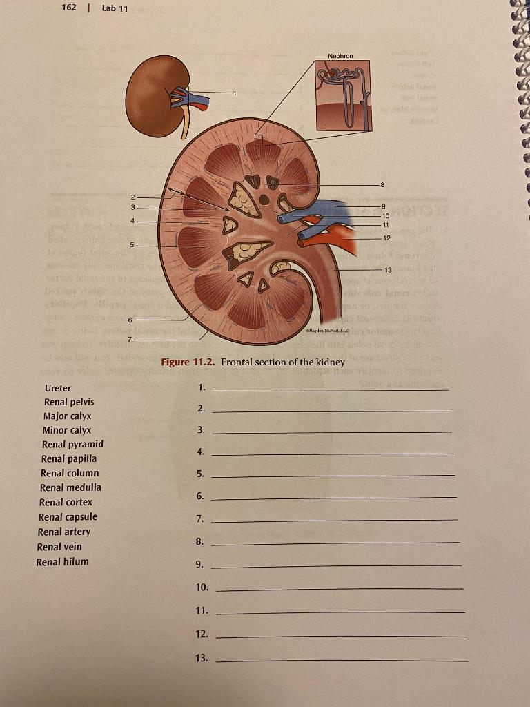
Solved SECTION 1: Overview of the Urinary System A. To help ...
Label the heart - Science Learning Hub In this interactive, you can label parts of the human heart. Drag and drop the text labels onto the boxes next to the diagram. Selecting or hovering over a box will highlight each area in the diagram. Aorta: The main artery carrying oxygenated blood to all parts of the body. Aorta. Vena cava: Carries deoxygenated blood from the body to the heart.

Comparative analysis of the combined petrosal and the ...
A Labeled Diagram of the Human Heart You Really Need to ... A Labeled Diagram of the Human Heart You Really Need to See. The heart, one of the most significant organs in the human body, is nothing but a muscular pump which pumps blood throughout the body. The human heart and its functions are truly fascinating. The heart, though small in size, performs highly significant functions that sustains human life.

Anterior Heart Frontal Section PART TWO Diagram | Quizlet
Three-dimensional ultrasound (frontal reformatted section ... Embed figure. Three-dimensional ultrasound (frontal reformatted section) of a bicornuate uterus. Note: The complete division of the uterine cavity and concave shape of the uterine fundus. Since ...

Malformations (Section 5) - Perinatal Neuropathology
Solved Chapter 11 The Cardiovascular System 181 6. Figure ... Figure 11-4 is a diagram of the frontal section of the heart. Follow the instructions below to complete this exercise. First, draw arrows to indicate the direction of blood flow through the heart. Draw the pathway of the oxygen-rich blood with red arrows, and trace the pathway of oxygen-poor blood with blue arrows. This problem has been solved!

Chapter 20, part 1 The Heart. - ppt video online download
33 Label This Anterior View Of The Human Heart - Labels ... Figure 4410 identify the features indicated on this anterior view of a frontal section of a human heart model using the terms provided. Correctly label the following anatomical features of the heart and thoracic cage correctly label the following vessels leading from and toward the anterior heart correctly label the following external anatomy ...

A two-stage framework for neural processing of biological ...
PDF Brain Anatomy - Wou BI 335 - Advanced Human Anatomy and Physiology Western Oregon University Figure 4: Mid-sagittal section of brain showing diencephalon (includes corpus callosum, fornix, and anterior commissure) Marieb & Hoehn (Human Anatomy and Physiology, 9th ed.) - Figure 12.10 Exercise 2: Utilize the model of the human brain to locate the following structures / landmarks for the

Mitral valve - Wikipedia
Anatomy of the normal fetal heart: The basis for ... (a) Anterior view of the thorax of a normal 22-week fetus, the heart located in the thoracic mediastinum. (b) Transversal section stained with hematoxylin and eosin from a human fetus at 20 weeks of development. Note the orientation of the heart in relation to the remaining structures. (c) Four chamber view at 25 weeks of gestation.

Origin and evolution of human speech: Emergence from a ...
PDF Brain Review and Wkst Answer Keys - Mayfield City School ... Created Date: 4/30/2013 4:05:46 PM

CS379C 2020 Class Discussion Notes
PDF The Cardiovascular 11 CHAPTER OUTLINE System The adult heart is shown in Figure 11.4 Note the thick ventricular walls, especially in the left ventricle. It is the left ventricle that must generate enough force to push blood throughout the body. The less muscular right ventricle pushes blood only to the nearby lungs.
Brain regions vulnerable and resistant to aging without ...
PDF Chapter 18 The Heart - Mrs. Ahrens' Science Site The figure below is a diagram of the frontal section of the heart. Follow the instructions below to complete this exercise, which considers both anatomical and physiological aspects of the heart. 1) Draw arrows to indicate the direction of blood flow through the heart.
Brain Sciences | Free Full-Text | Bi-Temporal Anodal ...
19.1 Heart Anatomy - Anatomy & Physiology Figure 19.1.1 - Position of the Heart in the Thorax: The heart is located within the thoracic cavity, medially between the lungs in the mediastinum. It is about the size of a fist, is broad at the top, and tapers toward the base. Shape and Size of the Heart
The metabolism and role of free fatty acids in key ...
NUMC 101 Module 2 Cardiovascular System Heart Lab Exercise ... Figure 11-4 is a diagram of the frontal section of the heart. Follow the instructions below to complete this exercise. First, draw arrows to indicate the direction of blood flow through the heart. Draw the pathway of the oxygen-rich blood with red arrows, and trace the pathway of the oxygen-poor blood with blue arrows.
Name: Period:
openstax.org › books › anatomy-and-physiology1.6 Anatomical Terminology - Anatomy and Physiology - OpenStax Figure 1.14 Planes of the Body The three planes most commonly used in anatomical and medical imaging are the sagittal, frontal (or coronal), and transverse plane. Body Cavities and Serous Membranes The body maintains its internal organization by means of membranes, sheaths, and other structures that separate compartments.
Homozygous silencing of T-box transcription factor EOMES ...
PDF Home - Buckeye Valley Created Date: 1/23/2014 12:31:42 PM

19.1 Heart Anatomy – Anatomy & Physiology

Dynamic Characteristics of Regional Flows around the Pyrénées ...

The Arcuate Fasciculus and language origins: Disentangling ...

The Cardiovascular System: The heart, vessels, and the ...

Electrocardiography in acute coronary syndromes | Advances in ...
Figure ll-4 Superior vena cava Left atrium 7. z. 4.

Frontal Section of the Heart Part 3 Diagram | Quizlet

Advances in the diagnosis of leukodystrophies | Future Neurology

Internal Features of the Heart (frontal section) Diagram ...
Cells | Free Full-Text | Potential Role of Intracranial Mast ...
Neurovascular Geography and Mapping the Consequences of Its ...
Frontiers | Sensorimotor Underpinnings of Mathematical ...

Frontal Section: Anatomy of heart Diagram | Quizlet

Heart Structure & Function - ppt download

An overview on stress neurobiology: Fundamental concepts and ...

The Arcuate Fasciculus and language origins: Disentangling ...

Lab Fig 3.4 Frontal section of the heart Diagram | Quizlet
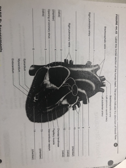
Solved FIGURE 44.15 Label this frontal section of the human ...

Chronological and Morphological Study of Heart Development in ...
0 Response to "36 figure 11-4 is a diagram of the frontal section of the heart"
Post a Comment